
Page 12 - KG_1 Booklet - 2019
P. 12
Kleiner et al Dovepress
biportal design of the delivery tool directed the slurry into radiographs revealing lucency around pedicle screws, lack of
the lateral areas of the prepared disk space. Once the disk graft incorporation, and/or greater than 2.5 degrees of motion
space was filled entirely, the site of insertion was inspected on flexion/extension radiographs. A visual analog scale
for any BG material. This material was excluded from the (VAS) for pain was obtained at each visit, and an Oswestry
final measurement to ensure an accurate calculation of BGD. Disability Index (ODI) was completed preoperatively and
Removal of the BGDT provided an unobscured path for 1.5 years postoperatively.
fusion cage application (Figure 6A and B).
A hollow polyether ether ketone, interbody fusion cage, Results
of the appropriate size was then placed into the disk space. There were 58 L4-5 disk spaces and 35 L5-S1 disk spaces
A minimally invasive, bilateral pedicle screw/rod system was evaluated. The average volumes of DMR from L4-5 and L5-S1
applied prior to wound closure. Average blood loss for the were 4.1±2.2 mL and 2.8±1.9 mL, respectively (Tables 1 and 2).
procedures was 127±75 mL. The P-value for the t-test was equal to 0.01, revealing a sig-
The volumes of DMR from L4-5 and L5-S1 were com- nificant difference in terms of DMR between L4-5 and L5-S1.
pared to one another, and the volumes of DMR were compared The comparison between DMR and BGD at L4-5 or at L5-S1
to the volume of BGD from the two disk spaces. A two-tailed demonstrated a significant difference (P,0.001).
Student’s t-test (t-test) was used to determine whether any BGD to L4-5 was 9.8±3.3 mL. At L5-S1, it was
significant difference existed between the volumes. The null 8.5±3.1 mL. The comparison between DMR and BGD at
hypothesis was that no significant difference existed between L4-5 or at L5-S1 demonstrated a significant difference
samples. Significance was set at P,0.05. (P,0.001). The P-value for the t-test was equal to 0.02,
In order to compare the volume of DMR during a T-LIF indicating a significant difference in BGD between L4-5 and
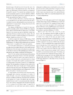
procedure with a complete, surgical diskectomy, the volume of L5-S1. The combined average was 9.2±3.0 mL. The amount
DMR during L5-S1 anterior lumbar diskectomy was measured of DMR compared to the amount of BGD to the disk space
volumetrically in an additional 29 consecutive patients. was not a 1:1 ratio, as would have been empirically expected.
The L5-S1 anterior diskectomy is a commonly performed At L4-5, the ratio was 3.1±2.1, and at L5-S1, it was 4.2±2.4
procedure allowing complete diskectomy with visualization and (Figure 8). This was statistically significant with a P-value
debridement of the entire space and represents an opportunity of 0.02. With respect to the entire study, the ratio of BGD
to calculate the volume of a thorough diskectomy relative to the relative to DMR revealed that on average 2.6±2.2 times as
anatomically and visually constrained T-LIF diskectomy. much BG was inserted into the disk space. This finding was
The L5-S1 disk was harvested and measured for patients even more dramatic with collapsed disk spaces where 1 mL
undergoing either anterior fusion or total disk replacement. of DMR led to an average of 6.6±0.9 mL of BGD. BGD was
The DMR consisted of anterior and posterior annulus as asymptotically related to the volume of DMR with 12.3 mL
well as complete nuclectomy and represented more tissue of BG being delivered to a disk where 8.0 mL of disk was
(in terms of the annuli) than would be typically removed in removed (Figure 9).
a T-LIF procedure. There were 29 anterior L5-S1 diskectomy
patients. The demographics and diagnosis were similar to the 10.0
T-LIF patients. There were 16 male and 13 female patients
with an average age of 56 years. The diagnosis for this group 7.5
of patients was spondylolisthesis and or degenerative osteoar- BG delivered is
almost 3x more than
thritis with central or lateral recess stenosis. the volume of disk
All study patients were followed with anterior/posterior Amount of material delivered (mL) 5.0 9.2±3.0 mL removed
radiographs and a physical examination at 4 weeks, 12
weeks, 26 weeks, 1 year, and 1.5 years postsurgery. Anterior/
posterior and flexion/extension radiographs were performed 2.5 3.6±2.1 mL
on all patients at 1.5 years. All patients who were symp-
tomatic between 1 and 1.5 years postoperatively underwent
0
flexion and extension radiographs and computed tomography BG delivered Disk removed
(CT) to evaluate for arthrodesis status. A suspicion of
Figure 8 BG delivered versus disk material removed from the L4-S1 disk spaces.
pseudoarthrosis was made based upon patient symptoms, Abbreviations: BG, bone graft; DMR, disk material removed.
110 submit your manuscript | www.dovepress.com Medical Devices: Evidence and Research 2016:9
Dovepress