
Page 8 - KG_1 Booklet - 2019
P. 8
Kleiner et al Dovepress
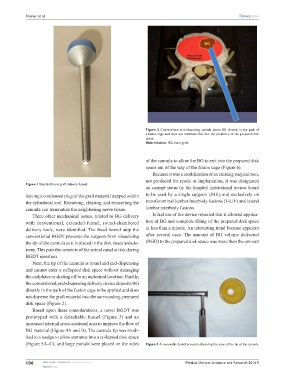
Figure 2 Conventional end-dispensing cannula ejects BG directly in the path of
a fusion cage and does not distribute BG into the periphery of the prepared disk
space.
Abbreviation: BG, bone graft.
of the cannula to allow the BG to exit into the prepared disk
space out of the way of the fusion cage (Figure 6).
Because it was a modification of an existing surgical tool,
not produced for resale or implantation, it was designated
Figure 1 Standard bone graft delivery funnel.
an exempt status by the hospital institutional review board
leaving a condensed plug of the graft material trapped within to be used by a single surgeon (JBK) and exclusively on
the cylindrical tool. Removing, clearing, and reinserting the transforaminal lumbar interbody fusions (T-LIF) and lateral
cannula can traumatize the neighboring nerve tissue. lumbar interbody fusions.
Three other mechanical issues, related to BG delivery Initial use of the device revealed that it allowed applica-
with conventional, extended-funnel, round-chambered tion of BG and complete filling of the prepared disk space
delivery tools, were identified. The fixed funnel atop the in less than a minute. An interesting trend became apparent
conventional BGDT prevents the surgeon from visualizing after several uses: The amount of BG volume delivered
the tip of the cannula as it is placed in the disk space annulo- (BGD) to the prepared disk space was more than the amount
tomy. This puts the contents of the spinal canal at risk during
BGDT insertion.
Next, the tip of the cannula is round and end-dispensing
and cannot enter a collapsed disk space without damaging
the endplates or skating off to an undesired location. Finally,
the conventional, end-dispensing delivery device deposits BG
directly in the path of the fusion cage to be applied and does
not disperse the graft material into the surrounding, prepared
disk space (Figure 2).
Based upon these considerations, a novel BGDT was
prototyped with a detachable funnel (Figure 3) and an
increased internal cross-sectional area to improve the flow of
BG material (Figure 4A and B). The cannula tip was modi-
fied to a wedge to allow entrance into a collapsed disk space
(Figure 5A–C), and large portals were placed on the sides Figure 3 A removable funnel prevents obscuring the view of the tip of the cannula.
106 submit your manuscript | www.dovepress.com Medical Devices: Evidence and Research 2016:9
Dovepress