Page 352 - Copper and Bronze in Art: Corrosion, Colorants, Getty Museum Conservation, By David Scott
P. 352

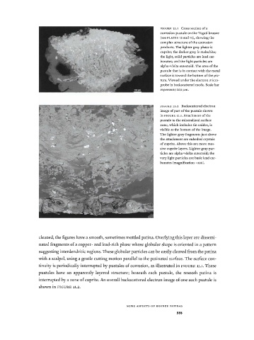
F I G U R E  íi.i  Cross section of a
                                                               corrosion pustule on the Togati bronze
                                                               (see  P L A T E S  73 and 75), showing the
                                                               complex structure  of the corrosion
                                                               products. The lighter gray phase is
                                                               cuprite; the darker gray is malachite;
                                                               the light, solid particles are lead car­
                                                               bonates; and the light particles are
                                                               alpha+delta eutectoid. The area of the
                                                               pustule that is in contact with the metal
                                                               surface is toward the bottom of the pic­
                                                               ture. Viewed under the electron micro-
                                                               probe in backscattered  mode. Scale bar
                                                               represents 100 μιη.


                                                               F I G U R E  11.2  Backscattered  electron
                                                               image of part of the pustule shown
                                                               in  F I G U R E  11.1. Attachment of the
                                                               pustule to the mineralized surface
                                                               zone, which includes tin oxides, is
                                                               visible at the bottom of the image.
                                                               The lighter gray fragments just above
                                                               the attachment  are euhedral  crystals
                                                               of cuprite. Above this are more mas­
                                                               sive cuprite layers. Lighter gray par­
                                                               ticles are alpha+delta eutectoid; the
                                                              very light particles are basic lead car­
                                                               bonates (magnification  χ 3 2 θ ) .


           cleaned, the figures have a smooth, sometimes mottled patina. Overlying this layer are dissemi­
           nated fragments of a copper- and lead-rich phase whose globular shape is oriented in a pattern
           suggesting interdendritic regions. These globular particles can be easily cleaved from the patina
           with  a scalpel, using a gentle cutting motion parallel to the patinated surface. The surface con­
           tinuity is periodically interrupted by pustules of corrosion,  as illustrated in FIGURE  11.1. These
           pustules  have  an  apparently  layered structure;  beneath  each  pustule,  the  smooth patina  is
           interrupted by a zone of cuprite. An overall backscattered electron image of one such pustule is
           shown in FIGURE  11.2.


                                                S O M E  A S P E C T S  O F  B R O N Z E  P A T I N A S
                                                                      335
   347   348   349   350   351   352   353   354   355   356   357