
Page 353 - Copper and Bronze in Art: Corrosion, Colorants, Getty Museum Conservation, By David Scott
P. 353
I SAMPLE DETAILS A core sample was taken through the hex
agonal network structure on the surface of the Roma bronze to examine the microstructure in
this area. PLATE 76 shows a cross section of this sample, taken through one of these hexagonal
surface features at xi30 magnification. Toward the top of the photomicrograph, an area of eutec
toid (the part of the structure examined through the hexagonal surface feature) is seen isolated
within the corrosion layer, showing that the copper-rich alpha phase of the bronze is preferen
tially corroded with preservation of eutectoid relicts. This microstructural study reveals that the
tin-enriched surface of the Roma bronze has not been formed by the corrosion of a deliberately
tinned surface layer. The part of the structure examined is not obviously dendritic; it has the
appearance of an annealed casting and could have been caused by the casting-on of the legs and
feet. The original dendritic and cored microstructure of the object could have been altered by
heating during the casting-on process to produce a microstructure more typical of annealed
bronzes. Changes in structure of this type have been seen in other works where casting-on has
been employed; an example is a dagger handle from Luristan, Iran (Scott 1991:94).
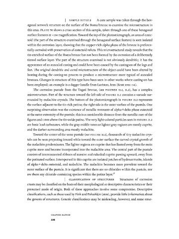
The corrosion pustule from the Togati bronze, (see FIGURES 11.1, 11.2), has a complex
microstructure. Part of the structure toward the left side of FIGURE 11.1 contains a vacuole sur
rounded by malachite crystals. The bottom of the photomicrograph in FIGURE 11.2 represents
the surface adjacent to the tin-rich patina; the right side is the outer surface of the pustule. One
surprising observation was the existence of metallic remnants of alpha+delta phase eutectoid
at the outer extremity of the pustule; this is a considerable distance from the metallic core of the
figures and 1 mm above the tin-oxide patina. The very light-colored particles seen in FIGURE 11.2
are basic lead carbonates, while the gray middle tones or lighter gray regions are mostly cuprite,
and the darker surrounding area mostly malachite.
Toward the center of the same pustule (see FIGURE 11.1), thousands of tiny malachite crys
tals can be seen projecting inward while toward the outer surface the curved crystal growth of
the malachite predominates. The lighter regions are cuprite that has floated away from the main
cuprite mass and become incorporated into the malachite area. The central part of the pustule
consists of interconnected ribbons of massive and euhedral cuprite passing upward, away from
the patinated surface. Interspersed in this cuprite are isolated patches of hydrocerrusite, islands
of alpha+delta eutectoid, and malachite. The malachite becomes more prevalent toward the
outer surface of the pustule. It is significant that there are no chlorides within the pustule, nor
are there any chloride-containing species within the patina layer. 17
I CLASSIFICATION OF STRUCTURES Structures of corrosion
crusts may be classified on the basis of their morphological or descriptive characteristics or their
presumed mode of origin. Both of these approaches involve some compromise. Descriptive
classifications, such as those used by Fink and Polushkin (i936), provide little information about
the genesis of structures. Genetic classifications may be misleading, however, and some struc-
C H A P T E R E L E V E N
336