
Page 403 - Copper and Bronze in Art: Corrosion, Colorants, Getty Museum Conservation, By David Scott
P. 403
system as a corrosion inhibitor, but this is not the case. The benzotriazole is added to act as a
uv absorber, so the loss of this compound from the coating implies that its stabilizer (BTA)
has evaporated. Voorhees (i988) describes in detail a six-step process to remove aged Incralac.
Bronze monuments were first treated with PMC protina solvent, which emulsified the lacquer
so that it could be scrubbed off with clean burlap. This was followed by a thorough rinsing, then
the procedure was repeated.
These studies show that, unfortunately, there is no easy way to combine reversibility
and exceptional protection in an organic coating. In the absence of a maintenance or moni
toring regime, there is no guarantee that any coating will perform as desired over a period of
several years.
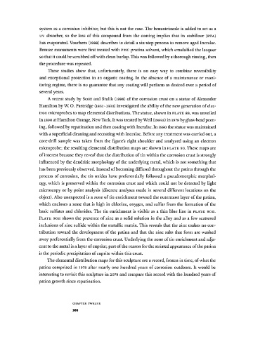
A recent study by Scott and Stulik (1998) of the corrosion crust on a statue of Alexander
Hamilton by W. O. Partridge (1861-1930) investigated the ability of the new generation of elec
tron microprobes to map elemental distributions. The statue, shown in PLATE 89, was unveiled
in 1890 at Hamilton Grange, New York. It was treated by Weil (i985a) in 1978 by glass-bead peen
ing, followed by repatination and then coating with Incralac. In 1980 the statue was maintained
with a superficial cleaning and recoating with Incralac. Before any treatment was carried out, a
core-drill sample was taken from the figure's right shoulder and analyzed using an electron
microprobe; the resulting elemental distribution maps are shown in PLATE 90. These maps are
of interest because they reveal that the distribution of tin within the corrosion crust is strongly
influenced by the dendritic morphology of the underlying metal, which is not something that
has been previously observed. Instead of becoming diffused throughout the patina through the
process of corrosion, the tin oxides have preferentially followed a pseudomorphic morphol
ogy, which is preserved within the corrosion crust and which could not be detected by light
microscopy or by point analysis (discrete analyses made in several different locations on the
object). Also unexpected is a zone of tin enrichment toward the outermost layer of the patina,
which encloses a zone that is high in chlorine, oxygen, and sulfur from the formation of the
basic sulfates and chlorides. The tin enrichment is visible as a thin blue line in PLATE 90B.
PLATE 90Ε shows the presence of zinc as a solid solution in the alloy and as a few scattered
inclusions of zinc sulfide within the metallic matrix. This reveals that the zinc makes no con
tribution toward the development of the patina and that the zinc salts that form are washed
away preferentially from the corrosion crust. Underlying the zone of tin enrichment and adja
cent to the metal is a layer of cuprite; part of the reason for the striated appearance of the patina
is the periodic precipitation of cuprite within this crust.
The elemental distribution maps for this sculpture are a record, frozen in time, of what the
patina comprised in 1978 after nearly one hundred years of corrosion outdoors. It would be
interesting to revisit this sculpture in 2078 and compare this record with the hundred years of
patina growth since repatination.
C H A P T E R T W E L V E
386