
Page 91 - Mesenchymal Stem Cell-Derived Exosomes as an Emerging Paradigm for Regenerative Therapy and Nano-Medicine
P. 91
CURRENT EYE RESEARCH 1363
Downloaded by [The UC Davis Libraries] at 16:12 05 October 2017
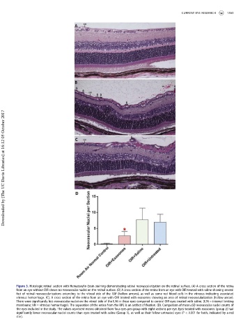
Figure 3. Histologic retinal section with Hematoxylin-Eosin staining demonstrating retinal neovascularization on the retinal surface. (A) A cross section of the retina
from an eye without OIR shows no neovascular nuclei on the retinal surface. (B) A cross section of the retina from an eye with OIR treated with saline showing several
foci of retinal neovascularizations extending to the vitreal side of the ILM (hollow arrows), as well as some red blood cells in the vitreous indicating associated
vitreous hemorrhage. (C). A cross section of the retina from an eye with OIR treated with exosomes showing an area of retinal neovascularization (hollow arrow).
There were significantly less neovascular nuclei on the vitreal side of the ILM in these eyes compared to control OIR eyes treated with saline. [ILM = internal limiting
membrane; VH = vitreous hemorrhage). The separation of the retina from the RPE is an artifact of fixation. (D). Comparison of mean±SD neovascular nuclei counts of
the eyes included in the study. The values represent means obtained from four eyes per group with eight sections per eye. Eyes treated with exosomes (group 2) had
significantly lower neovascular nuclei counts than eyes treated with saline (Group 1), as well as their fellow untreated eyes (P < 0.001 for both, indicated by a red
star).