
Page 90 - Mesenchymal Stem Cell-Derived Exosomes as an Emerging Paradigm for Regenerative Therapy and Nano-Medicine
P. 90
1362 E. MOISSEIEV ET AL.
Downloaded by [The UC Davis Libraries] at 16:12 05 October 2017
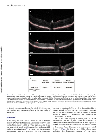
Figure 2. Composite OCT and phase-variance OCT angiography B-scan images of study eyes showing differences in retinal thickness in the three study groups. The
retina is notably thicker in the eye without OIR (A) when compared to the eye with OIR treated with saline (B) or exosomes (C). Red indicates vascular flow, and retinal
neovascularization is demonstrated by red on the retinal surface in eyes with OIR (B,C) (hollow arrows). The length of the vertical and horizontal arm of the L-shaped
scale at the lower left corner of each image represents 100 µm.(D) Mean ± SD of retinal thickness measurements in each study group. Both OIR groups (Groups 1 and 2)
had significantly reduced retinal thickness compared with the control group (Group 3); the retinal thickness was significantly reduced in saline-treated eyes (Group 1) in
comparison with exosome-treated eyes (Group 2) (p < 0.001 for all).
additional potential mechanism by which MSC exosomes murine eyes, that is, pvOCTA, as well as the traditional FA to
may mediate their protective effects in the OIR model of evaluate retinal perfusion in vivo. Furthermore, histologic
retinopathy. analysis was used to evaluate the effect of intravitreal admin-
istration of exosomes from human bone marrow MSCs in this
model of retinal ischemia.
Discussion
Both in vivo retinal imaging techniques, pvOCTA and FA,
In this study, we used a murine model of OIR to study the demonstrated that the degree of retinal ischemia and the
effect of intravitreal administration of exosomes derived from development of retinal neovascularization in eyes exposed to
human bone marrow MSCs on retinal ischemia. This model the OIR induction were reduced in eyes treated with intravi-
was used as it is a well-established, validated and quantifiable treal exosomes (Group 2) compared to saline treated controls
model for retinal ischemia. 20,24 We used a novel three-dimen- (Group 1) (Figure 1). The newer pvOCTA allows higher
sional in vivo retinal imaging system specifically designed for resolution three-dimensional imaging of the retinal