
Page 89 - Mesenchymal Stem Cell-Derived Exosomes as an Emerging Paradigm for Regenerative Therapy and Nano-Medicine
P. 89
CURRENT EYE RESEARCH 1361
Downloaded by [The UC Davis Libraries] at 16:12 05 October 2017
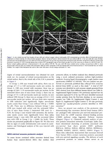
Figure 1. In vivo retinal vascular flow imaging of eyes with and without oxygen induced retinopathy (OIR) demonstrating protective effect of intravitreal exosome
treatment on retinal ischemia. (A-C) Fluorescein angiogram. Normal retinal perfusion is demonstrated in the eye without OIR (A). In the eyes with OIR, areas of retinal
ischemia and neovascularization are seen which are more pronounced in the eye that was treated with intravitreal saline (B) when compared to OIR eyes treated with
intravitreal exosomes (C). (D-F) Corresponding phase variance OCT angiography maps of the retinal vascular flow of the same eyes as shown in A (D), B (E) and C (F).
Normal retinal capillary perfusion is demonstrated in the eye without OIR (D). The eyes with OIR show marked retinal capillary non-perfusion which is more
pronounced in the eye that was treated with intravitreal saline (E) when compared to the eye treated with intravitreal exosomes (F). The horizontal lines are motion
artifacts. The length of the horizontal bar scale at the lower left corner of each image represents 0.17 mm.
degree of retinal neovascularization was obtained for each protective affects, we further analyzed data obtained previously
study eye. An example of retinal neovascularization on the using a novel, unbiased proteomics method, high-resolution
retinal surface, that is, the vitreal side of the ILM is presented isoelectric focusing liquid chromatography couple tandem mass
in Figure 3. spectrometry (HiRIEF LC-MS/MS). This new analysis was per-
In Group 1, OIR eyes treated with saline, there was an formed on previously published data collected from exosomes
average of 7.75 ± 3.68 neovascular nuclei per section. In derived from hMSC as used in this study. 17 A total of 1927
Group 2, OIR eyes treated with exosomes, there was an proteins were identified in each exosome sample generated from
average of 2.68 ± 1.35 neovascular nuclei per section. In the MSCs derived from three different human donors (see Table S1,
untreated fellow eyes of mice from Group 2, there was an Supplemental Digital Content 1.We previously reported that the
average of 7.0 ± 2.48 neovascular nuclei per section. In eyes exosomes expressed 92 of the top 100 most identified exosomal
without OIR (Group 3), there was an average of 0.12 ± 0.33 marker proteins from the ExoCarta database in our exosome
neovascular nuclei per section. All eyes from mice exposed samples (see Table S2, Supplemental Digital Content 2, and
to OIR induction had significantly higher neovascular Figure S1, Supplemental Digital Content 3). We also previously
nuclei counts than Group 3 eyes without OIR (p < 0.0001 reported the vascular-protective proteins identified in MSC
for all). There was no difference between Group 1 eyes exosomes. 17
(OIR eyes treated with saline) and the untreated fellow Here, we present new analysis of the proteomic data
eyes of mice from Group 2 (untreated OIR eyes) (p = demonstrating that exosomes derived from human MSCs
0.35). In Group 2 eyes treated with exosomes, the neovas- are packaged with numerous pro-survival-associated pro-
cular nuclei counts were significantly lower than saline teins from the cAMP response element-binding protein
treated Group 1 eyes with OIR and the untreated fellow (CREB) pathway using Ingenuity Pathway Analysis
eyes with OIR in Group 2 (p < 0.0001 for both). These (Qiagen) (Figure 4). Clustered network analysis of pro-
results are presented graphically in Figure 3D. We also note tein–protein interaction networks (CytoScape, Spike data-
that no signs of ocular inflammation were noted on exam- base) determined clustering of proteins associated with
ination and histological analysis of the eyes injected with pro-survival heat shock protein (HSP) pathways:
exosomes. HSPA1A, HSPA4, HSPA5, HSPA8, HSPA9, HSP90AA1,
HSPB90, HSPBP1, HSPD1, HSPG2, HSPH1 (Figure 5).
These data collectively demonstrate that exosomes derived
hMSCs-derived exosome proteomic analysis
from human MSCs contain numerous proteins associated
To assess factors contained within exosomes derived from with pro-survival signaling cascades. The delivery of such
human bone marrow-derived MSCs that mediate their prosurvival proteins to retinal tissues serves as an