
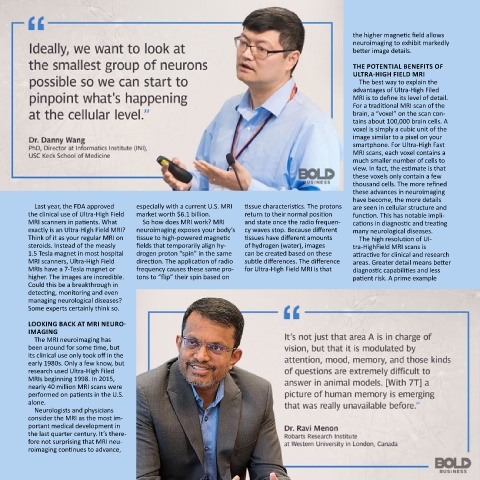
Page 26 - BB Neuromedicine Highlights 2019
P. 26
the higher magnetic field allows
                                                                                           neuroimaging to exhibit markedly
                                                                                           better image details.

                                                                                           The POTeNTIal BeNeFITs OF
                                                                                           ulTra-hIgh FIelD mrI
                                                                                             The best way to explain the
                                                                                           advantages of Ultra-High Filed
                                                                                           MRI is to define its level of detail.
                                                                                           For a traditional MRI scan of the
                                                                                           brain, a “voxel” on the scan con-
                                                                                           tains about 100,000 brain cells. A
                                                                                           voxel is simply a cubic unit of the
                                                                                           image similar to a pixel on your
                                                                                           smartphone. For Ultra-High Fast
                                                                                           MRI scans, each voxel contains a
                                                                                           much smaller number of cells to
                                                                                           view. In fact, the estimate is that
                                                                                           these voxels only contain a few
                                                                                           thousand cells. The more refined
                                                                                           these advances in neuroimaging
                                                                                           have become, the more details
          Last year, the FDA approved   especially with a current U.S. MRI   tissue characteristics. The protons   are seen in cellular structure and
        the clinical use of Ultra-High Field   market worth $6.1 billion.    return to their normal position   function. This has notable impli-
        MRI scanners in patients. What   So how does MRI work? MRI   and state once the radio frequen-  cations in diagnostic and treating
        exactly is an Ultra-High Field MRI?   neuroimaging exposes your body’s   cy waves stop. Because different   many neurological diseases.
        Think of it as your regular MRI on   tissue to high-powered magnetic   tissues have different amounts   The high resolution of Ul-
        steroids. instead of the measly   fields that temporarily align hy-  of hydrogen (water), images   tra-HighField MRI scans is
        1.5 Tesla magnet in most hospital   drogen proton “spin” in the same   can be created based on these   attractive for clinical and research
        MRI scanners, Ultra-High Field   direction. The application of radio   subtle differences. The difference   areas. Greater detail means better
        MRIs have a 7-Tesla magnet or   frequency causes these same pro-  for Ultra-High Field MRI is that   diagnostic capabilities and less
        higher. The images are incredible.   tons to “flip” their spin based on            patient risk. A prime example
        Could this be a breakthrough in
        detecting, monitoring and even
        managing neurological diseases?
        Some experts certainly think so.

        lOOkINg Back aT mrI NeurO-
        ImagINg
          the mRi neuroimaging has
        been around for some time, but
        its clinical use only took off in the
        early 1980s. Only a few know, but
        research used Ultra-High Filed
        MRIs beginning 1998. In 2015,
        nearly 40 million MRI scans were
        performed on patients in the U.S.
        alone.
          Neurologists and physicians
        consider the mRi as the most im-
        portant medical development in
        the last quarter century. it’s there-
        fore not surprising that MRI neu-
        roimaging continues to advance,