
Page 252 - Medicine and Surgery
P. 252
P1: KPE
BLUK007-06 BLUK007-Kendall May 25, 2005 18:6 Char Count= 0
248 Chapter 6: Genitourinary system
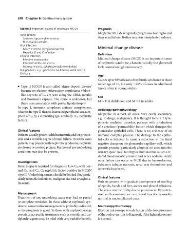
Table 6.9 Important causes of secondary MCGN Prognosis
Idiopathic MCGN is typically progressive leading to end
Autoimmune
Systemic lupus erythematosus stagerenalfailure.Itoftenrecursintransplantedkidneys.
Rheumatoid arthritis
Viral infection
Mixed essential cryoglobulinaemia Minimal change disease
Hepatitis B and C infection
Chronic infection Definition
Infective endocarditis Minimal change disease (MCD) is an important cause
Infected ventricular shunts of nephrotic syndrome, characteristically the glomeruli
Leprosy, malaria, schistosomiasis (worldwide) look normal on light microscopy.
Malignancies, e.g. lymphoma, leukaemia, renal cell Ca
Cirrhosis
Age
Causesupto90%ofcasesofnephroticsyndromeinthose
under age of 10, but only ∼20% of cases in adulthood
Type II MCGN is also called ‘dense deposit disease’
(more often in young adults).
because on electron microscopy, continuous ribbon-
like deposits of C 3 are seen along the GBM, tubules
Sex
and Bowman’s capsule. The cause is unknown, but
M > Finchildhood, and M = F in adults.
there is an association with partial lipodystrophy.
In type I, immune complexes activate complement,
Aetiology/pathophysiology
whereas in type II there is increased peripheral consum-
Idiopathic in almost all cases. Very rarely secondary,
ption of C 3 by a circulating IgG antibody (C 3 nephritic
e.g. to drugs, malignancy. It is thought to beaTlym-
factor).
phocyte mediated disorder, perhaps with production
of a cytokine (permeability factor) which damages the
Clinical features glomerular epithelial cells. There is no evidence of an
Patientsusuallypresentwithhaematuriaand/orprotein- immune complex process. The damage to the epithe-
uria and a variable degree of renal failure. In severe cases lial cells is believed to cause a reduction in the fixed
patientsmaypresentwithnephroticsyndrome,nephritic negative charge on the glomerular capillary wall, which
syndrome or a mixed picture. Features of any underlying permits protein (particularly albumin) to cross into the
condition may also be present. urinary space. Resultant hypoalbuminaemia causes a re-
duced blood oncotic pressure and hence oedema. Acute
renal failure can occur in MCD due to hypovolaemia,
Investigations
ischaemic tubular necrosis, renal vein thrombosis and
Renal biopsy is required for diagnosis. Low C 3 ,with nor-
interstitial nephritis.
mal C 1q and C 4 .C 3 nephritic factor positive in MCGN
type II. Underlying causes should be looked for, partic-
Clinical features
ularly treatable infections, malignancies and cryoglobu-
Patients present with gradual development of swelling
linaemia.
of eyelids, hands and feet, ascites and pleural effusions.
The urine may be frothy due to proteinuria. Hyperten-
Management sion and haematuria are rare. Renal function is usually
Treatment of any underlying cause may lead to partial normal in uncomplicated cases.
or complete remission. In those without nephrotic syn-
drome, conservative management is probably indicated, Macroscopy/microscopy
as the prognosis is good. In those with nephrotic-range Electron microscopy reveals fusion of the foot processes
proteinuria, specific treatments such as steroids and an- ofthepodocytes,thisisdiagnosticifthelightmicroscopy
tiplatelet agents may be tried with very variable benefit. is normal.