
Page 340 - Medicine and Surgery
P. 340
P1: FAW
BLUK007-07 BLUK007-Kendall May 25, 2005 18:18 Char Count= 0
336 Chapter 7: Nervous system
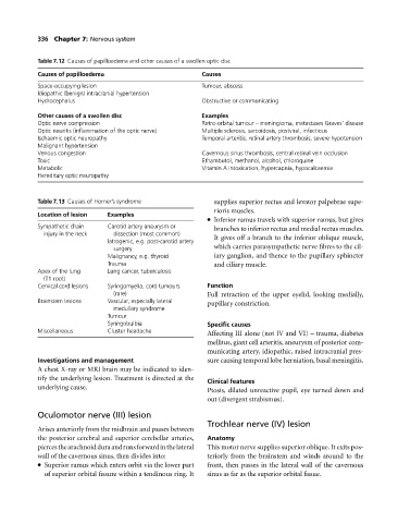
Table 7.12 Causes of papilloedema and other causes of a swollen optic disc
Causes of papilloedema Causes
Space-occupying lesion Tumour, abscess
Idiopathic (benign) intracranial hypertension
Hydrocephalus Obstructive or communicating
Other causes of a swollen disc Examples
Optic nerve compression Retro-orbital tumour – meningioma, metastases Graves’ disease
Optic neuritis (inflammation of the optic nerve) Multiple sclerosis, sarcoidosis, postviral, infectious
Ischaemic optic neuropathy Temporal arteritis, retinal artery thrombosis, severe hypotension
Malignant hypertension
Venous congestion Cavernous sinus thrombosis, central retinal vein occlusion
Toxic Ethambutol, methanol, alcohol, chloroquine
Metabolic Vitamin A intoxication, hypercapnia, hypocalcaemia
Hereditary optic neuropathy
Table 7.13 Causes of Horner’s syndrome supplies superior rectus and levator palpebrae supe-
rioris muscles.
Location of lesion Examples
Inferior ramus travels with superior ramus, but gives
Sympathetic chain Carotid artery aneurysm or branches to inferior rectus and medial rectus muscles.
injury in the neck dissection (most common) It gives offabranchto the inferior oblique muscle,
Iatrogenic, e.g. post-carotid artery
surgery which carries parasympathetic nerve fibres to the cil-
Malignancy, e.g. thyroid iary ganglion, and thence to the pupillary sphincter
Trauma and ciliary muscle.
Apex of the lung Lung cancer, tuberculosis
(T1 root)
Cervical cord lesions Syringomyelia, cord tumours Function
(rare) Full retraction of the upper eyelid, looking medially,
Brainstem lesions Vascular, especially lateral pupillary constriction.
medullary syndrome
Tumour
Syringobulbia Specific causes
Miscellaneous Cluster headache Affecting III alone (not IV and VI) – trauma, diabetes
mellitus, giant cell arteritis, aneurysm of posterior com-
municating artery, idiopathic, raised intracranial pres-
Investigations and management sure causing temporal lobe herniation, basal meningitis.
Achest X-ray or MRI brain may be indicated to iden-
tify the underlying lesion. Treatment is directed at the
Clinical features
underlying cause.
Ptosis, dilated unreactive pupil, eye turned down and
out (divergent strabismus).
Oculomotor nerve (III) lesion
Trochlear nerve (IV) lesion
Arises anteriorly from the midbrain and passes between
the posterior cerebral and superior cerebellar arteries, Anatomy
piercesthearachnoidduraandrunsforwardinthelateral This motor nerve supplies superior oblique. It exits pos-
wall of the cavernous sinus, then divides into: teriorly from the brainstem and winds around to the
Superior ramuswhich enters orbit via the lower part front, then passes in the lateral wall of the cavernous
of superior orbital fissure within a tendinous ring. It sinus as far as the superior orbital fissue.