
Page 341 - Medicine and Surgery
P. 341
P1: FAW
BLUK007-07 BLUK007-Kendall May 25, 2005 18:18 Char Count= 0
Chapter 7: Disorders of cranial and peripheral nerves 337
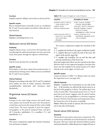
Function Table 7.14 Causes of trigeminal nerve (V) lesions
Supplies superior oblique (moves the eye down and in).
Location Examples of causes
Brainstem (pons, medulla, Infarct, tumour, multiple
Specific causes
upper cervical cord) sclerosis, syringobulbia
Rare as isolated lesion, Generally occurs as a combined Posterior fossa Aneurysm, tumour, meningitis
III, IV and VI nerve palsies (see below) when the eye is Petrous temporal bone Acoustic neuroma, trauma
also intorted. (fracture of bone),
meningioma or other tumour
Cavernous sinus (only V 1 Tumour, cavernous sinus
Clinical features
branch of V and usually thrombosis, aneurysm of the
Diplopia on looking down or in. III, IV and VI also internal carotid.
affected)
Abducent nerve (VI) lesion
The sensory components supply the sensation of the
Anatomy face:
Supplies lateral rectus. It exits from the brainstem and 1 V 1 supplies the forehead, the upper eyelid and eyeball.
runs through the subarachnoid space into the cavernous 2 V 2 supplies the lower eyelid, the side of the nose, the
sinus, then passes through the superior orbital fissue. upper teeth and the upper lip.
3 V 3 supplies the mandible, the ear and the skin and
Function mucous membranes of the lower jaw.
Lateral rectus deviates the eye laterally. Pain and temperature fibres are also carried on the three
divisions back to the trigeminal ganglion, but then dive
Specific causes down into the medulla to the spinal nucleus of V which
Particularly at risk from raised intracranial pressure or extends as far as the upper cervical cord.
traumaduetoitslongcourse.Oftenoccursasacombined
III, IV and VI nerve palsies Specific causes
Causes are shown in Table 7.14. Herpes zoster can infect
the trigeminal nerve (see page 326).
Clinical features
Diplopia on looking to the side. III, IV and VI combined
Clinical features
nerve palsies are seen in stroke, tumours, Wernicke’s
Sensory:Complete loss of sensation on one side of the
encephalopathy, aneurysms and cavernous sinus
face – if all branches are affected the lesion must be at
thrombosis.
the level of the ganglion or above. The earliest sign is loss
of the corneal reflex. Dissociated sensory loss (i.e. loss of
Trigeminal nerve (V) lesion pain but touch intact) suggests only the spinal nucleus
is affected, e.g. by syringobulbia or a foramen magnum
Anatomy
tumour. If touch is lost, but pain and temperature intact,
Emerges as two roots (large sensory and small motor
the lesion has to be in the pons or medulla.
root), passes out forwards the pons into the posterior
Motor:When the mouth is opened, the lower jaw devi-
cranial fossa and across the temporal lobe in the middle
ates to the side of the lesion.
cranial fossa. The nerve expands to form the trigemi-
nal ganglion, at the petrous temporal bone, and gives
Facial nerve (VII) lesions
off 3 branches: ophthalmic (V 1 ), maxillary (V 2 ) and
mandibular (V 3 ). Anatomy
Thefacialnervehasmotorandsensorycomponents.The
Function motor nerve cell bodies are in the facial nerve nucleus in
The motor components supply the muscles of mastica- the pons. The nerve enters the internal auditory meatus
tion and tensor tympani. and passes laterally within the petrous temporal bone to