
Page 381 - Medicine and Surgery
P. 381
P1: KTX
BLUK007-08 BLUK007-Kendall May 12, 2005 19:48 Char Count= 0
Chapter 8: Vasculitis 377
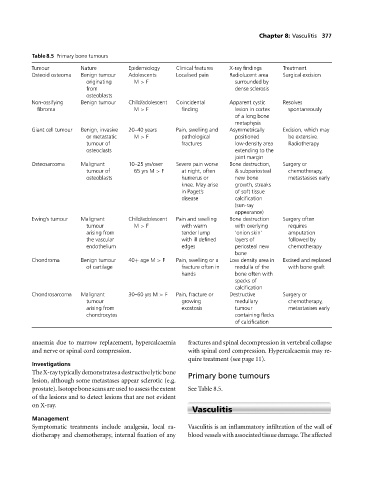
Table 8.5 Primary bone tumours
Tumour Nature Epidemiology Clinical features X-ray findings Treatment
Osteoid osteoma Benign tumour Adolescents Localised pain Radiolucent area Surgical excision
originating M > F surrounded by
from dense sclerosis
osteoblasts
Non-ossifying Benign tumour Child/adolescent Coincidental Apparent cystic Resolves
fibroma M > F finding lesion in cortex spontaneously
of a long bone
metaphysis
Giant cell tumour Benign, invasive 20–40 years Pain, swelling and Asymmetrically Excision, which may
or metastatic M > F pathological positioned be extensive.
tumour of fractures low-density area Radiotherapy
osteoclasts extending to the
joint margin
Osteosarcoma Malignant 10–25 yrs/over Severe pain worse Bone destruction, Surgery or
tumour of 65 yrs M > F at night, often & subperiosteal chemotherapy,
osteoblasts humerus or new bone metastasises early
knee. May arise growth, streaks
in Paget’s of soft tissue
disease calcification
(sun-ray
appearance)
Ewing’s tumour Malignant Child/adolescent Pain and swelling Bone destruction Surgery often
tumour M > F with warm with overlying requires
arising from tender lump ‘onion skin’ amputation
the vascular with ill defined layers of followed by
endothelium edges periosteal new chemotherapy
bone
Chondroma Benign tumour 40+ age M > F Pain, swelling or a Low density area in Excised and replaced
of cartilage fracture often in medulla of the with bone graft
hands bone often with
specks of
calcification
Chondrosarcoma Malignant 30–60 yrs M > F Pain, fracture or Destructive Surgery or
tumour growing medullary chemotherapy,
arising from exostosis tumour metastasises early
chondrocytes containing flecks
of calcification
anaemia due to marrow replacement, hypercalcaemia fractures and spinal decompression in vertebral collapse
and nerve or spinal cord compression. with spinal cord compression. Hypercalcaemia may re-
quire treatment (see page 11).
Investigations
TheX-raytypicallydemonstratesadestructivelyticbone
Primary bone tumours
lesion, although some metastases appear sclerotic (e.g.
prostate). Isotope bone scans are used to assesstheextent See Table 8.5.
of the lesions and to detect lesions that are not evident
on X-ray.
Vasculitis
Management
Symptomatic treatments include analgesia, local ra- Vasculitis is an inflammatory infiltration of the wall of
diotherapy and chemotherapy, internal fixation of any blood vessels with associated tissue damage. The affected