
Page 4 - UNIT 3
P. 4
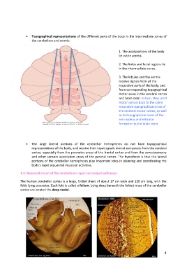
Topographical representations of the different parts of the body in the intermediate zones of
the cerebellum and vermis:
1. The axial portions of the body
lie in the vermis.
1
2. The limbs and facial regions lie
in the intermediate zones.
3. The lobules and the vermis
receive signals from all the
respective parts of the body, and
2 2 from corresponding topographical
motor areas in the cerebral cortex
and brain stem. In turn, they send
motor signals back to the same
respective topographical areas of
the cerebral motor cortex, as well
as to topographical areas of the
red nucleus and reticular
formation in the brain stem.
The large lateral portions of the cerebellar hemispheres do not have topographical
representations of the body, and receive their input signals almost exclusively from the cerebral
cortex, especially from the premotor areas of the frontal cortex and from the somatosensory
and other sensory association areas of the parietal cortex. The hypothesis is that the lateral
portions of the cerebellar hemispheres play important roles in planning and coordinating the
body's rapid sequential muscular activities.
1.3. Neuronal circuit of the cerebellum: input and output pathways.
The human cerebellar cortex is a large, folded sheet of about 17 cm wide and 120 cm long, with the
folds lying crosswise. Each fold is called a folium. Lying deep beneath the folded mass of the cerebellar
cortex are located the deep nuclei.
4