
Page 13 - PR 2014 2016 10 Materials and Nanotechnology
P. 13
Materials and Nanotechnology | Progress Report 289
sonication and centrifugation, rGO powders peak at 26.3° corresponding to the (002) plane.
are oven-dried and deagglomerated. The appli- On the conversion to graphene oxide (GO),
cability of these powders has been evaluated this peak gradually shifts to a lower angle
for electrocatalyst support for polymeric fuel (2θ= 9.1°). Wide peak between 15° and 35°
cell, supercapacitor electrodes and zirconia might be related to the formation of reduced
bioceramics reinforcement. graphene oxide since methanol has shown a
reducing capability towards GO. After reduc-
tion of GO, the (002) plane shifted towards a
higher angle value (2θ= 24.5°) due to removal
of some oxygen functional groups. A repre-
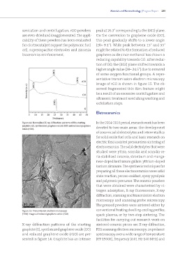
sentative transmission electron microscopy
image of rGO is shown in figure 15. The ob-
served fragmented thin film feature might
be a result of an excessive centrifugation and
ultrasonic treatment used along washing and
exfoliation steps.
Eletroceramics
Figure 14: Normalized X-ray diffraction patterns of the starting In the 2014-2016 period, research work has been
graphite (G), synthesized graphene oxide (GO) and reduced graphene devoted to two main areas: the development
oxide (rGO).
of ceramic solid electrolytes and intermetallics
for solid oxide fuel cells and basic research on
electric field assisted pressureless sintering of
electroceramics. The solid electrolytes that were
studied were yttria, scandia and scandia-ce-
ria stabilized zirconia, strontium and manga-
nese-doped lanthanum gallate, yttrium-doped
barium zirconate. The synthesis techniques for
preparing all these electroceramics were solid
state reaction, peroxo-oxidant, spray pyrolysis
and polymeric precursor. The ceramic powders
that were obtained were characterized by ni-
trogen adsorption, X-ray fluorescence, X-ray
diffraction, scanning and transmission electron
microscopy and scanning probe microscopy.
The pressed powders were sintered either by
conventional heating dwelling-cooling profiles,
Figure 15: Transmission electron microscopy
(TEM) images of reduced graphene oxide (rGO) spark plasma, or by two-step sintering. The
facilities for carrying out research work on
X-ray diffraction patterns of the starting sintered ceramic pieces are X-ray diffraction,
graphite (G), synthesized graphene oxide (GO) FEG scanning electron microscopy, impedance
and reduced graphene oxide (rGO) are pre- spectroscopy over a wide range of temperature
sented in figure 14. Graphite has an intense (RT-1500K), frequency (0.01 Hz-140 MHz) and