
Page 7 - CBAC Newsletter 2013
P. 7
modeling metaBoliSm-exCitation CouPling:
k aS the gateway
atP
By Jonathan r. Silva and Colin g. niCholS
Background
Among channels that regulate excitability, the ATP-sensitive K channel (K ) is unique because it provides a direct link
+
ATP
to cellular metabolism. Thus, channels open to provide outward, repolarizing K current in response to altered con-
+
centrations of intracellular nucleotides. In the beta-cells of the pancreas, reduced [ATP] and increased ADP and AMP
i
caused by lower plasma glucose concentration suppresses Ca channel-driven electrical bursts. The lower bursting
2+
rate results in less Ca entry, which in turn lowers [Ca ] and the rate of insulin secretion. Consequently, mutations
2+
2+
i
that cause constitutively open K lead to profound neonatal diabetes mellitus (NDM) in both mice and humans, due
ATP
to a suppression of bursting and therefore a lack of insulin secretion (Koster, Marshall et al. 2000; Gloyn, Pearson et
al. 2004). Surprisingly, NDM patients have not been found to exhibit any cardiac phenotype, even though K channels
ATP
are present at very high density in the mycocyte sarcolemmal membrane (Noma 1983). The existence of unique car-
diac and pancreatic phenotypes has been attributed to heterogeneity in the molecular composition of K ATP in different
tissues. However, several recent publications, including the discovery of a link between K ATP mutations and the Early
Repolarization Syndrome (ERS) (Haissaguerre, Chatel et al. 2009), have called the uniqueness of the cardiac K chan-
ATP
nel molecular composition into question. Here, we will discuss these findings and future experimental and theoretical
directions that could clarify the consequences of heterogeneous K expression.
ATP
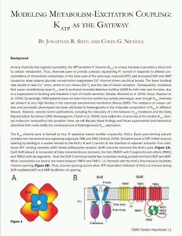
The K ATP channel pore is formed by four K -selective inward rectifier α-subunits, Kir6.x. Each pore-forming subunit
+
contains two transmembrane-spanning segments TM1 and TM2 (Nichols 2006). Elevated levels of ATP inhibit channel
opening by binding to a pocket formed by the Kir6.x N and C termini at the interface of adjacent subunits. Four addi-
tional ATP- binding cassette (ABC) family-sulfonylurea receptor (SUR) subunits surround the Kir6.x pore (Figure 1A).
Each SUR subunit is composed of three transmembrane domains, the first (TMD0) with 5 segments and others (TMD1
and TMD2) with six segments. Near the SUR C-terminus reside the nucleotide binding pockets that bind ADP and AMP.
When nucleotides are bound, the linker between TMD0 and TMD1, L0, interacts with the Kir6.x N-terminus to facilitate
channel opening (Figure 1B). Thus, channel opening occurs when ATP-dependent inhibition of Kir6.x is relieved during
SUR-mediated ADP and AMP facilitation of opening.
Figure 1
CBAC Center Heartbeat |1