
Page 128 - Zoo Animal Learning and Training
P. 128
Chapter 13: Surgical Treatment of Skull Tumors 127
A B
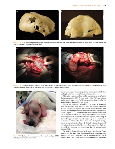
Figure 13.12 (A, B) A custom‐printed sterilizable three‐dimensional printed skull cap can be used intraoperatively to help contour the titanium plate in a
manner that closely resembles the normal skull.
A B
Figure 13.13 (A) Placement of an intraoperatively contoured titanium mesh that has been fixed in place with small bone screws. (B) Appearance of the mesh
following closure of any remaining temporalis muscle prior to subcutaneous and skin closure.
increased procedure time, and a potential increased risk of infection
leading to empyema or need to remove the implant.
PMMA has been successfully used to reconstruct large defects in
the canine calvarium [20]. A preformed molded PMMA prosthetic
implant can be used or the PMMA prosthesis can be sculpted at the
time of surgery (Figures 13.9 and 13.10).
Surgical titanium mesh is available in a variety of styles and
sizes. The standard mesh is 0.6 mm thick and is easily contoured
by hand. Equipment required includes the mesh, associated self‐
drilling/self‐tapping screws, specialized screwdriver, and fine wire
cutters (Figure 13.11). Reconstruction mesh benders and cutters
can be purchased but are not a requirement to use this implant.
Following removal of the affected bone segment, contouring of
the mesh is performed to reconstruct the region of bone that has
been removed. The mesh is first cut to size and then gently manip-
ulated to reconstruct the defect. If desired, a three‐dimensional
printed sterilizable model of the normal contour of the skull can
be used for planning and contouring during reconstruction
(Figure 13.12).
The mesh is then fixed to the skull with self‐drilling/self‐tap-
ping screws that have been premeasured based on preoperative
Figure 13.14 Postoperative appearance of the patient in Figure 13.13 imaging (Figure 13.13). Predrilling is recommended if the bone is
following reconstruction and closure. greater than 5 mm thick, which is uncommon in veterinary