
Page 132 - Zoo Animal Learning and Training
P. 132
Chapter 14: Shunt Placement and Marsupialization in Treatment of Hydrocephalus and Quadrigeminal Diverticula 131
A B
3 2
1
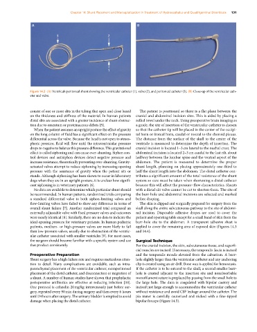
Figure 14.2 (A) Ventriculoperitoneal shunt showing the ventricular catheter (1), valve (2), and peritoneal catheter (3). (B) Close‐up of the ventricular cath-
eter and valve.
consist of one or more slits in the tubing that open and close based The patient is positioned so there is a flat plane between the
on the thickness and stiffness of the material. In human patients cranial and abdominal incision sites. This is aided by placing a
distal slits are associated with a greater incidence of shunt obstruc- rolled towel under the neck. Using preoperative brain imaging as
tion due to omentum or proteinaceous debris [5]. a guide, the site of insertion of the ventricular catheter is chosen
When the patient assumes an upright posture the effect of gravity so that the catheter tip will be placed in the center of the occipi-
on the long column of fluid has a significant effect on the pressure tal horn or frontal horn, caudal or rostral to the choroid plexus.
differential across the valve. Because the head is not open to atmos- The distance from the surface of the skull to the center of the
pheric pressure, fluid will flow until the intraventricular pressure ventricle is measured to determine the depth of insertion. The
drops to negative to balance this pressure difference. This gravitational cranial incision is located 1–3 cm lateral to the nuchal crest. The
effect is called siphoning and can cause over‐shunting. Siphon con- abdominal incision is located 2–3 cm caudal to the last rib, about
trol devices and antisiphon devices detect negative pressure and halfway between the lumbar spine and the ventral aspect of the
increase resistance, theoretically preventing over‐shunting. Gravity‐ abdomen. The patient is measured to determine the proper
actuated valves attempt to reduce siphoning by increasing opening shunt length, planning on placing approximately one‐third to
pressure with the assistance of gravity when the patient sits or half the shunt length into the abdomen. The distal catheter con-
stands. Although siphoning has been shown to occur in laboratory tributes a significant amount of the total resistance of the shunt
dogs when they are in an upright posture, it is unclear how signifi- system so care must be taken when shortening a distal catheter
cant siphoning is in veterinary patients [6]. because this will affect the pressure–flow characteristics. Shunts
No data are available to determine which particular shunt should with a distal slit valve cannot be cut to shorten them. The site of
be recommended. In human patients, randomized trials comparing the burr‐hole and abdominal incisions are selected and marked
a standard differential valve to both siphon‐limiting valves and before draping.
flow‐limiting valves have failed to show any difference in terms of The skin is clipped and surgically prepared for surgery from the
overall shunt failure [7]. Another randomized trial compared an skull along the entire subcutaneous pathway to the site of abdomi-
externally adjustable valve with fixed pressure valves and outcomes nal incision. Disposable adhesive drapes are used to cover the
were nearly identical [8]. Similarly, there are no data to indicate the patient and operating table except for a small band of skin from the
ideal opening pressure for veterinary patients. In human pediatric burr‐hole site to the abdomen. A transparent adhesive sheet is
patients, medium‐ or high‐pressure valves are more likely to fail applied to cover the remaining area of exposed skin (Figures 14.3
than low‐pressure valves, usually due to obstruction of the ventric- and 14.4).
ular catheter associated with smaller ventricles [9]. For most cases,
the surgeon should become familiar with a specific system and use Surgical Technique
that product consistently. For the cranial incision, the skin, subcutaneous tissue, and superfi-
cial muscles are incised. If necessary, the temporalis fascia is incised
Preoperative Preparation and the temporalis muscle elevated from the calvarium. A burr‐
Shunt surgery has a high failure rate and requires meticulous atten- hole slightly larger than the ventricular catheter and any anchoring
tion to detail. Many complications are avoidable, such as intra- clip is created using an air drill. Bone wax is applied for hemostasis.
parenchymal placement of the ventricular catheter, extraperitoneal If the catheter is to be sutured to the skull, a second smaller burr‐
placement of the distal catheter, and disconnection or migration of hole is created adjacent to the insertion site and nonabsorbable
a shunt. A number of human studies have shown that prophylactic monofilament suture is preplaced by passing from the small hole to
perioperative antibiotics are effective at reducing infection [10]. the large hole. The dura is coagulated with bipolar cautery and
One protocol is cefazolin 20 mg/kg intravenously just before sur- incised just large enough to accommodate the ventricular catheter
gery, repeated every 90 min during surgery and then every 6 hours without resistance and avoid CSF leakage around the catheter. The
until 24 hours after surgery. The urinary bladder is emptied to avoid pia mater is carefully cauterized and nicked with a fine‐tipped
damage when placing the distal catheter. bipolar forceps (Figure 14.5).