
Page 136 - Zoo Animal Learning and Training
P. 136
Chapter 14: Shunt Placement and Marsupialization in Treatment of Hydrocephalus and Quadrigeminal Diverticula 135
A B
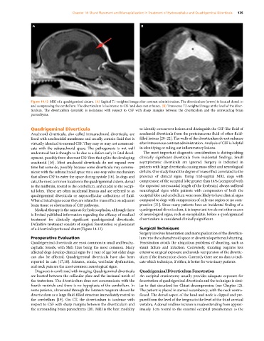
Figure 14.12 MRI of a quadrigeminal cistern. (A) Sagittal T1‐weighted image after contrast administration. The diverticulum (arrow) is located dorsal to
and compressing the cerebellum. The diverticulum is isointense to CSF and does not enhance. (B) Transverse T2‐weighted image at the level of the diver-
ticulum. The diverticulum (asterisk) is isointense with respect to CSF with sharp margins between the diverticulum and the surrounding brain
parenchyma.
Quadrigeminal Diverticula to identify concurrent lesions and distinguish the CSF‐like fluid of
Arachnoid diverticula, also called intraarachnoid diverticula, are arachnoid diverticula from the proteinaceous fluid of other fluid‐
lined with arachnoidal membrane and usually contain fluid that is filled lesions [20–22]. The walls of the diverticulum do not enhance
virtually identical to normal CSF. They may or may not communi- after intravenous contrast administration. Analysis of CSF is helpful
cate with the subarachnoid space. The pathogenesis is not well in identifying or ruling out inflammatory lesions.
understood but is thought to be due to a defect early in fetal devel- The most important diagnostic consideration is distinguishing
opment, possibly from aberrant CSF flow that splits the developing clinically significant diverticula from incidental findings. Small
arachnoid [16]. Most arachnoid diverticula do not expand over asymptomatic diverticula are ignored. Surgery is indicated in
time but some do, possibly because some diverticula may commu- patients with large diverticula causing mass effect and neurological
nicate with the subarachnoid space via a one‐way valve mechanism deficits. One study found the degree of mass effect correlated to the
that allows CSF to enter the space during systole [16]. In dogs and presence of clinical signs. Using mid‐sagittal MRI, dogs with
cats, the most common location is the quadrigeminal cistern, dorsal compression of the occipital lobe greater than 14% (compared with
to the midbrain, rostral to the cerebellum, and caudal to the occipi- the expected rostrocaudal length of the forebrain) always suffered
tal lobes. These are often incidental lesions and are referred to as neurological signs while patients with compression of both the
quadrigeminal diverticula or supracollicular collections of fluid. occipital lobe and cerebellum were more likely to have clinical signs
When clinical signs occur they are related to mass effect on adjacent compared to dogs with compression of only one region or no com-
brain tissue or obstruction of CSF pathways. pression [21]. Since many patients have an incidental finding of a
Medical therapy is the same as for hydrocephalus, although there quadrigeminal diverticulum, it is important to rule out other causes
is limited published information regarding the efficacy of medical of neurological signs, such as encephalitis, before a quadrigeminal
treatment for clinically significant quadrigeminal diverticula. diverticulum is considered clinically significant.
Definitive treatment consists of surgical fenestration or placement
of a diverticuloperitoneal shunt (Figure 14.12). Surgical Techniques
Surgery involves fenestration and marsupialization of the diverticu-
Preoperative Evaluation lum into the subarachnoid space or diverticuloperitoneal shunting.
Quadrigeminal diverticula are most common in small and brachy- Fenestration avoids the ubiquitous problems of shunting, such as
cephalic breeds, with Shih Tzus being the most common. Many shunt failure and infection. Conversely, shunting requires less
affected dogs develop clinical signs by 1 year of age but older dogs extensive surgical exposure and avoids reexpansion of the divertic-
can also be affected. Quadrigeminal diverticula have also been ulum if the fenestration closes. Currently there are no data to indi-
reported in cats [17,18]. Seizures, ataxia, vestibular dysfunction, cate which technique, if either, is better for veterinary patients.
and neck pain are the most common neurological signs.
Diagnosis is confirmed with imaging. Quadrigeminal diverticula Quadrigeminal Diverticulum Fenestration
are located between the collicular plate and the incisural notch of An occipital craniectomy usually provides adequate exposure for
the tentorium. The diverticulum does not communicate with the fenestration of quadrigeminal diverticula and the technique is simi-
fourth ventricle and there is no hypoplasia of the cerebellum. In lar to that described for Chiari decompression (see Chapter 12).
some patients, ultrasound through the foramen magnum shows the The patient is placed in sternal recumbency, with the neck ventro-
diverticulum as a large fluid‐filled structure immediately rostral to flexed. The dorsal aspect of the head and neck is clipped and pre-
the cerebellum [19]. On CT, the diverticulum is isodense with pared from the level of the bregma to the level of the third cervical
respect to CSF with sharp margins between the diverticulum and vertebra. A dorsal midline incision is made extending from approx-
the surrounding brain parenchyma [20]. MRI is the best modality imately 1 cm rostral to the external occipital protuberance to the