
Page 141 - Zoo Animal Learning and Training
P. 141
142 Section III: Spinal Procedures
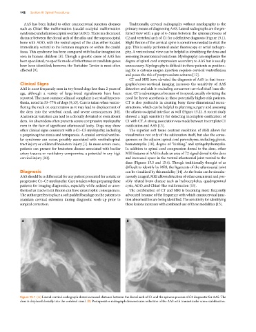
AAS has been linked to other craniocervical junction diseases Traditionally, cervical radiography without myelography is the
such as Chiari‐like malformation (caudal occipital malformation primary means of diagnosing AAS. Lateral radiographs are the pre-
syndrome) and atlantooccipital overlap (AOO). There is a decreased ferred view with a gap of 4–5 mm between the spinous process of
distance between the dorsal arch of the atlas and the supraoccipital C2 and vertebral arch of C1 for a definitive diagnosis (Figure 15.1).
bone with AOO, with the rostral aspect of the atlas either located Slight flexion of the cervical spine is sometimes needed to elicit the
immediately ventral to the foramen magnum or within the caudal gap. This is safely performed under fluoroscopy or serial radiogra-
fossa. This syndrome has been compared with basilar invagination phy. A ventrodorsal view can be helpful in identifying the dens and
seen in human children [8]. Though a genetic cause of AAS has assessing its anatomical variations. Myelography can emphasize the
been speculated, no specific mode of inheritance or candidate genes degree of spinal cord compression secondary to AAS but is usually
have been identified; however, the Yorkshire Terrier is most often unnecessary. Myelography is difficult in these patients as position-
affected [9]. ing for a cisterna magna injection requires cervical ventroflexion
and poses the risk of postprocedure seizures [12].
CT and MRI have elevated the diagnosis of AAS in that tomo-
Clinical Signs graphic/cross‐sectional imaging increases the sensitivity of AAS
AAS is most frequently seen in toy‐breed dogs less than 2 years of detection and aids in excluding concurrent cervical/skull base dis-
age, although a variety of large‐breed signalments have been ease. CT is advantageous because of its speed, usually obviating the
reported. The most common clinical complaint is cervical hyperes- need for heavy anesthesia in these potentially higher‐risk patients.
thesia, noted in 53–77% of dogs [9,10]. Care is taken when ventro- CT is also preferable in creating bony three‐dimensional recon-
flexing the neck on examination as it may lead to displacement of structions, which can be helpful in planning surgery and assessing
the dens into the vertebral canal, and severe consequences [10]. the atlanto-occipital interface as well (Figure 15.2). A recent study
Anatomical variation can lead to a dorsally deviated or even absent showed a high sensitivity for detecting incomplete ossification of
dens. An absent dens often prevents severe compressive myelopathy C1 with CT. A strong association was made between incomplete C1
even in the face of significant atlantoaxial laxity. Dogs may show ossification and AAS [13].
other clinical signs consistent with a C1–C5 myelopathy, including The superior soft tissue contrast resolution of MRI allows for
a proprioceptive ataxia and tetraparesis. A cranial cervical vestibu- visualization not only of the subluxation itself, but also the conse-
lar syndrome can occur, possibly associated with vestibulospinal quences on the adjacent spinal cord parenchyma, including gliosis,
tract injury or collateral brainstem injury [1]. In more severe cases, hematomyelia [10], degree of “kinking,” and syringohydromyelia.
patients can present for brainstem disease associated with basilar In addition to spinal cord compression dorsal to the dens, other
artery trauma or ventilatory compromise, a potential in any high MRI features of AAS include an area of T2 signal dorsal to the dens
cervical injury [10]. and increased space in the ventral atlantoaxial joint ventral to the
dens (Figures 15.3 and 15.4). Though traditionally thought of as
difficult to identify by MRI, the ligaments of the atlantoaxial joint
Diagnosis can be visualized by this modality [14]. As the brain can be simulta-
AAS should be a differential for any patient presented for a static or neously imaged, MRI allows detection of other concurrent and pos-
progressive C1–C5 myelopathy. Care is taken when preparing these sibly related brain disease such as hydrocephalus, quadrigeminal
patients for imaging diagnostics, especially while sedated or anes- cysts, AOO, and Chiari‐like malformation [15].
thetized as inadvertent flexion can have catastrophic consequences. The combination of CT and MRI is becoming more frequently
The author prefers to place a soft padded bandage on the patients to advocated because of the frequency with which craniocervical junc-
maintain cervical extension during diagnostic work‐up prior to tion abnormalities are being identified. The sensitivity for identifying
surgical correction. these lesions increases with combined use of these modalities [15].
A B
Figure 15.1 (A) Lateral cervical radiograph shows increased distance between the dorsal arch of C1 and the spinous process of C2 diagnostic for AAS. The
dens is displaced dorsally into the vertebral canal. (B) Postoperative radiograph demonstrates reduction of the AAS with transarticular screw stabilization.