
Page 142 - Zoo Animal Learning and Training
P. 142
Chapter 15: Atlantoaxial Subluxation 143
Medical Management Surgical Procedures
The two primary indications for medical management are patient Surgical stabilization of AAS can be classified as dorsal and ventral
age and financial constraints for the client. As AAS patients are approaches [1]. The ventral approaches are more commonly
most frequently toy‐breed animals, those younger than 6–8 months utilized and are reported to have a higher rate of success [9].
are often not considered good candidates for surgery because of size
challenges and lack of boney mineralization. Surgical correction is
often delayed until there is radiographic evidence of physis closure A
at the vertebral endplate.
In the author’s experience, the frequent replacement of the splint
compounded with possible complications associated with the coap-
tation is often financially comparable to the surgical procedure
itself. Havig et al. [16] reported on the medical management of
AAS, citing a 10 of 16 case success with external coaptation when
used for 1 month, particularly when the clinical signs were for less
than 30 days. Ventral splinting is easier, with a contoured rigid plate
made of fiberglass or orthoplast maintained in position by a modi-
fied Robert Jones type bandage (Figure 15.5).
B
Figure 15.3 (A) Cervical neutral and (B) flexed positioning can be per-
formed in the MRI unit. Note on the flexed view the increased space
between the ventral body of C1 and the dens as well as the adjacent dorsal
Figure 15.2 Three‐dimensional CT of an atlantoaxial subluxation. compressive myelopathy.
A B
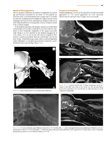
Figure 15.4 A 5‐year‐old female spayed Pekinese presents for a C1–C5 myelopathy. (A) Lateral radiograph demonstrates atlantoaxial subluxation and con-
genital fusion of the C2–C3 vertebral bodies. (B) MRI demonstrates the atlantoaxial subluxation and congenital C2–C3 fusion plus a severe compressive
myelopathy secondary to intervertebral disc disease at C3–C4.