
Page 143 - Zoo Animal Learning and Training
P. 143
144 Section III: Spinal Procedures
A B
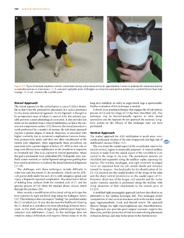
Figure 15.5 Types of external coaptation used for conservative therapy and postoperatively for approximately 6 weeks to maintain the atlantoaxial joint in
an extended position to allow fusion. (A) A contoured rigid plate made of fiberglass or orthoplast maintained in position by a modified Robert Jones type
bandage. (B) A soft, commercially available splint.
Dorsal Approach long‐term stabilizer, its safety in larger‐breed dogs is questionable.
The dorsal approach to the cervical spine in cases of AAS is desira- Further evaluation of this technique is needed.
ble in that it has the potential for placement of a replica prosthesis A dorsal cross‐pinning technique that engages the dorsal spinous
for the dorsal atlantoaxial ligament. As this ligament is thought to process of C2 and the wings of C1 has been described [22]. This
be an important cause of failure in cases of AAS, this solution pos- technique may be biomechanically superior to other dorsal
sibly provides a more physiological correction. It also provides less approaches and the implants do not approach the neuraxis. Long‐
strain on the implants than a ventral stabilization, as this is the ten- term studies on the efficacy of this technique have not been
sion not compression surface [17]. However, the dorsal procedure is performed.
rarely performed for a number of reasons: the soft tissue approach
requires a greater degree of muscle dissection, is associated with Ventral Approach
higher morbidity due to incisional complications (seroma forma- The ventral approach for AAS stabilization is much more com-
tion, postoperative pain), and does not allow visualization of the monly performed because of the ease of approach and high rate of
ventral joint alignment. Most importantly, these procedures are stabilization success (Video 15.1).
associated with a greater degree of failure (37–49%) as they rely on The area from the caudal aspect of the mandibular rami to the
long‐term fibrous tissue stabilization as the prosthesis is suspected caudal cervical region is aseptically prepared. A ventral midline
to eventually fail. This is in contrast to ventral approaches, where incision is made from the caudal aspect of the mandible to 3 cm
the goal is permanent bony arthrodesis [9]. Orthopedic wire, syn- caudal to the wings of the atlas. The sternohyoid muscles are
thetic suture material, or nuchal ligament autogenous grafting have identified and separated along the midline raphe, exposing the
been used as prostheses to replicate the dorsal atlantoaxial ligament trachea. The trachea, esophagus, and right recurrent laryngeal
[17–19]. nerve are separated from the left carotid sheath and retracted
The challenges with these techniques include difficulty in toward the surgeon. Two landmarks for the identification of the
reduction and placement of the prosthesis, which can be diffi- C1–C2 junction are the caudal borders of the wings of the atlas
cult particularly under the arch of C1, with iatrogenic spinal cord and the sharp ventral prominence on the caudal aspect of C1.
injury a frequently reported complication. Also described is failure Periosteal dissection of the longus colli muscles and the rectus
of holding bony surfaces (both the vertebral arch of C1 and the capitis ventralis muscles is performed along the midline with
spinous process of C2) when the implant shears (cheese wires) sharp dissection of their attachments to the ventral spine of
through the purchase [20]. C2 [23].
More recently, a modification of the dorsal wiring technique has A modified right parasaggital approach has been described as an
been reported in eight toy‐breed dogs using the Kishigami device alternative to the midline technique [24]. This technique avoids
[21]. This technique does not require “feeding” the prosthesis under manipulation of vital cervical structures such as the trachea, esoph-
the C1 vertebral arch. It may also decrease the likelihood of tearing agus, vagosympathetic trunk, and thyroid vessels. The approach
the C1 dorsal arch and allows for more physiological lateral move- dissects though the right sternocephalicus and sternothyroideus.
ment. In this study, seven of eight dogs achieved good long‐term This approach also provides good surgical exposure, requires less
reduction and stabilization (1 year). As this technique does not dissection, provides protection of vital structures during placement
intend to induce arthrodesis and requires fibrous tissue to be the of fixation devices, and may better preserve the thyroid artery.