
Page 135 - Zoo Animal Learning and Training
P. 135
134 Section II: Intracranial Procedures
Postoperative Management Very small ventricles allow the brain to almost completely fill the
Pain control is provided by injectable analgesics that are transi- intracranial space, which decreases the ability to compensate for
tioned to oral medication. Preoperative antibiotics are occasionally transient increases in intracranial volume. Episodes of pain can
continued for several days but prolonged antibiotic therapy is not occur after shunting in dogs and may be similar to slit ventricle
indicated for uncomplicated cases. Any preoperative antiseizure syndrome in human patients [12].
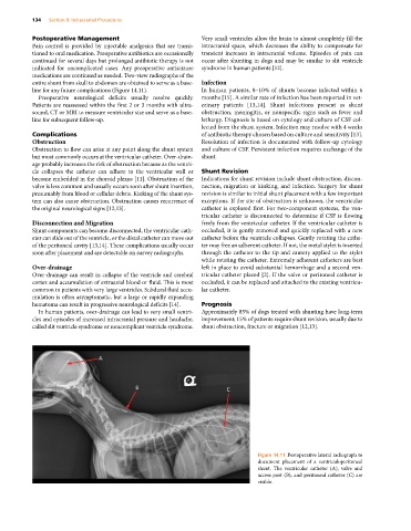
medications are continued as needed. Two‐view radiographs of the
entire shunt from skull to abdomen are obtained to serve as a base- Infection
line for any future complications (Figure 14.11). In human patients, 8–10% of shunts become infected within 6
Preoperative neurological deficits usually resolve quickly. months [15]. A similar rate of infection has been reported in vet-
Patients are reassessed within the first 2 or 3 months with ultra- erinary patients [13,14]. Shunt infections present as shunt
sound, CT or MRI to measure ventricular size and serve as a base- obstruction, meningitis, or nonspecific signs such as fever and
line for subsequent follow‐up. lethargy. Diagnosis is based on cytology and culture of CSF col-
lected from the shunt system. Infection may resolve with 4 weeks
Complications of antibiotic therapy chosen based on culture and sensitivity [13].
Obstruction Resolution of infection is documented with follow‐up cytology
Obstruction to flow can arise at any point along the shunt system and culture of CSF. Persistent infection requires exchange of the
but most commonly occurs at the ventricular catheter. Over‐drain- shunt.
age probably increases the risk of obstruction because as the ventri-
cle collapses the catheter can adhere to the ventricular wall or Shunt Revision
become embedded in the choroid plexus [11]. Obstruction of the Indications for shunt revision include shunt obstruction, discon-
valve is less common and usually occurs soon after shunt insertion, nection, migration or kinking, and infection. Surgery for shunt
presumably from blood or cellular debris. Kinking of the shunt sys- revision is similar to initial shunt placement with a few important
tem can also cause obstruction. Obstruction causes recurrence of exceptions. If the site of obstruction is unknown, the ventricular
the original neurological signs [12,13]. catheter is explored first. For two‐component systems, the ven-
tricular catheter is disconnected to determine if CSF is flowing
Disconnection and Migration freely from the ventricular catheter. If the ventricular catheter is
Shunt components can become disconnected, the ventricular cath- occluded, it is gently removed and quickly replaced with a new
eter can slide out of the ventricle, or the distal catheter can move out catheter before the ventricle collapses. Gently rotating the cathe-
of the peritoneal cavity [13,14]. These complications usually occur ter may free an adherent catheter. If not, the metal stylet is inserted
soon after placement and are detectable on survey radiographs. through the catheter to the tip and cautery applied to the stylet
while rotating the catheter. Extremely adherent catheters are best
Over‐drainage left in place to avoid substantial hemorrhage and a second ven-
Over‐drainage can result in collapse of the ventricle and cerebral tricular catheter placed [2]. If the valve or peritoneal catheter is
cortex and accumulation of extraaxial blood or fluid. This is most occluded, it can be replaced and attached to the existing ventricu-
common in patients with very large ventricles. Subdural fluid accu- lar catheter.
mulation is often asymptomatic, but a large or rapidly expanding
hematoma can result in progressive neurological deficits [14]. Prognosis
In human patients, over‐drainage can lead to very small ventri- Approximately 85% of dogs treated with shunting have long‐term
cles and episodes of increased intracranial pressure and headache, improvement; 15% of patients require shunt revision, usually due to
called slit ventricle syndrome or noncompliant ventricle syndrome. shunt obstruction, fracture or migration [12,13].
Figure 14.11 Postoperative lateral radiograph to
document placement of a ventriculoperitoneal
shunt. The ventricular catheter (A), valve and
access port (B), and peritoneal catheter (C) are
visible.