
Page 1197 - Equine Clinical Medicine, Surgery and Reproduction, 2nd Edition
P. 1197
1172 CHAPTER 11
VetBooks.ir Table 11.7 Common aetiologies of conjunctivitis 11.81
• Bacterial
• Streptococcus equi, Actinobacillus spp., Chlamydia,
Rhodococcus spp., Moraxella equi, Leptospira spp.
• Viral
• Adenovirus, equine herpesvirus (EHV)-1
(rhinopneumonitis), EHV-2 (cytomegalovirus), EHV-4?,
equine infectious anaemia, equine viral arteritis, equine
influenza type A2, parainfluenza, African horse sickness
(reovirus)
• Mycotic
• Histoplasma capsulatum var. farciminosum (also called
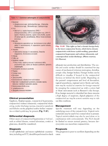
called H. farciminosum), H. capsulatum, sporotrichosis, Fig. 11.81 This right eye had a chronic foreign body
blastomycosis
• Parasitic in the lower conjunctival fornix, which led to chronic
• Habronema muscae, H. microstoma, Draschia conjunctivitis with lower eyelid swelling, generalised
megastoma, Onchocerca cervicalis conjunctival hyperaemia and oedema (chemosis), and
• Allergic a mucopurulent ocular discharge. (Photo courtesy
• Follicular GA Munroe)
• Systemic causes
• Pneumonia, equine protozoal myeloencephalitis,
polyneuritis equi, vestibular disease syndrome, epizootic adequate tear production and distribution. The eye-
lymphangitis lids and ocular surface should be examined for any
• Eosinophilic keratoconjunctivitis
• Trauma causes of mechanical irritation (entropion, distichia,
• Foreign body ectopic cilia, foreign bodies). Foreign bodies can be
• Entropion difficult to visualise if located in the conjunctival
• Dacryocystitis fornices or behind the third eyelid. Depending on
• Environmental irritants/chemical irritation the patient’s temperament and level of discomfort,
• Neonatal maladjustment syndrome, neonatal sepsis, topical anaesthesia, regional nerve blocks and seda-
immune-mediated haemolytic anemia
• Keratoconjunctivitis sicca tion may be needed to examine for foreign material
• Lymphosarcoma by sweeping the conjunctival sac with a cotton bud
• Idiopathic/immune-mediated or blunt instrument such as Bennett’s cilia forceps.
If no foreign material is identified but there remains
a high degree of suspicion, digital palpation of the
Clinical presentation conjunctival sac may be a more sensitive way to rule
Epiphora, blepharospasm, conjunctival hyperaemia, out this condition.
conjunctival oedema (chemosis), conjunctival thick-
ening, ocular discharge (serous, mucoid or purulent) Management
and follicles on the palpebral and/or bulbar conjunc- Specific treatment will vary depending on the
tiva or third eyelid may be present (Fig. 11.81). underlying cause of the conjunctivitis. Topical corti-
costeroids may be used if no corneal ulcer is present.
Differential diagnosis Topical antimicrobials may also be used alone or in
Other causes of conjunctival hyperaemia or ‘red eye’, combination with corticosteroids. The NLS should
such as orbital disease, corneal ulceration, uveitis or be flushed to ensure normal patency, because dac-
glaucoma, should be considered. ryocystitis can exacerbate conjunctivitis.
Diagnosis Prognosis
A full ophthalmic and neuro-ophthalmic examina- The prognosis is poor to excellent depending on the
tion, including an STT, should be performed to check cause.