
Page 1195 - Equine Clinical Medicine, Surgery and Reproduction, 2nd Edition
P. 1195
1170 CHAPTER 11
VetBooks.ir 11.77 11.78
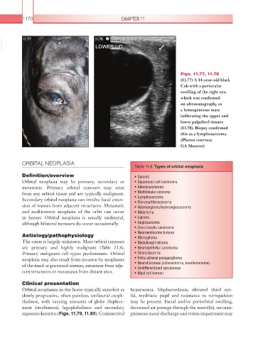
Figs. 11.77, 11.78
(11.77) A 14-year-old black
Cob with a periocular
swelling of the right eye,
which was confirmed
on ultrasonography as
a homogeneous mass
infiltrating the upper and
lower palpebral tissues
(11.78). Biopsy confirmed
this as a lymphosarcoma.
(Photos courtesy
GA Munroe)
ORBITAL NEOPLASIA
Table 11.6 Types of orbital neoplasia
Definition/overview • Sarcoid
Orbital neoplasia may be primary, secondary or • Squamous cell carcinoma
metastatic. Primary orbital tumours may arise • Adenocarcinoma
from any orbital tissue and are typically malignant. • Multilobular osteoma
Secondary orbital neoplasia can involve local exten- • Lymphosarcoma
• Fibroma/fibrosarcoma
sion of masses from adjacent structures. Metastatic • Haemangioma/haemangiosarcoma
and multicentric neoplasia of the orbit can occur • Melanoma
in horses. Orbital neoplasia is usually unilateral, • Lipoma
although bilateral tumours do occur occasionally. • Angiosarcoma
• Granulocytic carcinoma
Aetiology/pathophysiology • Neuroendocrine tumour
• Microglioma
The cause is largely unknown. Most orbital tumours • Medulloepithelioma
are primary and highly malignant (Table 11.6). • Neuroepithelial carcinoma
Primary malignant cell types predominate. Orbital • Osteoclastoma
neoplasia may also result from invasion by neoplasms • Extra-adrenal paraganglioma
of the nasal or paranasal sinuses, extension from adja- • Neurofibromas (schwannoma, neurilemmoma)
• Undifferentiated carcinomas
cent structures or metastases from distant sites. • Mast cell tumour
Clinical presentation
Orbital neoplasms in the horse typically manifest as hyperaemia, blepharoedema, elevated third eye-
slowly progressive, often painless, unilateral exoph- lid, mydriatic pupil and resistance to retropulsion
thalmos, with varying amounts of globe displace- may be present. Facial and/or periorbital swelling,
ment (strabismus), lagophthalmos and secondary decreased air passage through the nostril(s), serosan-
exposure keratitis (Figs. 11.79, 11.80). Conjunctival guineous nasal discharge and vision impairment may