
Page 1200 - Equine Clinical Medicine, Surgery and Reproduction, 2nd Edition
P. 1200
Eyes 1175
VetBooks.ir elastase by the corneal epithelial cells, leucocytes and vary considerably, but can include blepharospasm,
photophobia, epiphora, serous to mucopurulent
certain microbial organisms (especially Pseudomonas
and beta-haemolytic streptococci) results in sudden,
sis, conjunctivitis, corneal oedema, variable corneal
rapid degeneration of collagen and other compo- ocular discharge, conjunctival hyperaemia, chemo-
nents of the stroma, inducing corneal liquefaction or neovascularisation (superficial and/or deep), white to
keratomalacia (corneal ‘melting’). Keratomalacia can grey to brown plaque adhered to the corneal surface
lead to globe rupture in less than 12 hours if it is not (fungal infections) (Figs. 11.83, 11.84), interstitial
controlled. keratitis and white–yellow or grey gelatinous cor-
The presence of anterior uveitis secondary to cor- neal opacity or exudates (stromal necrosis/liquefac-
neal disease is common in horses. Anterior uveitis tion or keratomalacia). Signs of corneal ulcers can be
can lead to scarring and/or blockage of the ICA and/ quite subtle, especially in sick or hospitalised foals
or uveoscleral outflow pathway and cause an eleva- because their corneas are significantly less sensitive
tion in the IOP or glaucoma. than those of normal foals or adult horses. Clinical
Predisposing factors for corneal ulceration signs of secondary anterior uveitis, ranging in sever-
include prolonged topical antimicrobial, cortico- ity, are also commonly seen in horses with corneal
steroid or corticosteroid/antimicrobial combination disease (i.e. miosis, aqueous flare, hypopyon). Other
drugs, which may inhibit the growth of normal bac- associated complications of corneal ulceration
teria and predispose to mycotic infection. include scarring, pigmentation, anterior and poste-
rior synechiae, cataract formation, endophthalmitis,
Clinical presentation phthisis bulbi and blindness.
Corneal ulcers can range in appearance from simple,
superficial breaks or abrasions in the corneal epithe- Differential diagnosis
lium not visible to the naked eye, to deep stromal A corneal facet (an ulcer that has re-epithelialised),
ulcers, to full-thickness corneal perforations with stromal abscess, uveitis, glaucoma and other causes
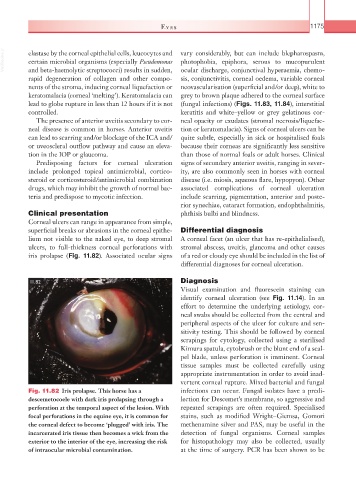
iris prolapse (Fig. 11.82). Associated ocular signs of a red or cloudy eye should be included in the list of
differential diagnoses for corneal ulceration.
11.82 Diagnosis
Visual examination and fluorescein staining can
identify corneal ulceration (see Fig. 11.14). In an
effort to determine the underlying aetiology, cor-
neal swabs should be collected from the central and
peripheral aspects of the ulcer for culture and sen-
sitivity testing. This should be followed by corneal
scrapings for cytology, collected using a sterilised
Kimura spatula, cytobrush or the blunt end of a scal-
pel blade, unless perforation is imminent. Corneal
tissue samples must be collected carefully using
appropriate instrumentation in order to avoid inad-
vertent corneal rupture. Mixed bacterial and fungal
Fig. 11.82 Iris prolapse. This horse has a infections can occur. Fungal isolates have a predi-
descemetocoele with dark iris prolapsing through a lection for Descemet’s membrane, so aggressive and
perforation at the temporal aspect of the lesion. With repeated scrapings are often required. Specialised
focal perforations in the equine eye, it is common for stains, such as modified Wright–Giemsa, Gomori
the corneal defect to become ‘plugged’ with iris. The methenamine silver and PAS, may be useful in the
incarcerated iris tissue then becomes a wick from the detection of fungal organisms. Corneal samples
exterior to the interior of the eye, increasing the risk for histopathology may also be collected, usually
of intraocular microbial contamination. at the time of surgery. PCR has been shown to be