
Page 1207 - Equine Clinical Medicine, Surgery and Reproduction, 2nd Edition
P. 1207
1182 CHAPTER 11
VetBooks.ir Aetiology/pathophysiology 11.91
As with corneal ulcers, corneal abscesses may be
bacterial, fungal, or sterile in origin (see Table 11.8).
Corneal stromal abscesses develop following focal
trauma to the cornea that allows opportunis-
tic pathogens and debris into the stroma beneath
the corneal epithelium. Subsequent healing or
re- epithelialisation of this ulcer or epithelial micro-
puncture forms a barrier that protects the bacteria
or fungi from topically administered antimicro-
bial medications, sealing in the microorganisms
and allowing ongoing infection. Alternatively, the
initial treatment may kill the microorganisms, but 11.92
subsequent release of toxins by dying bacteria and
fungi, as well as degenerating leucocytes, continues
the stimulus for abscessation. Topical antimicrobial/
corticosteroid combination therapy can predispose
horses to developing corneal abscessation. Fungi
appear to have a predilection for the deep corneal
stroma and/or Descemet’s membrane. Concurrent
anterior uveitis occurs, due to corneal sensory nerve
irritation (oculopupillary reflex). Anterior uveitis is
the main cause of posterior synechia, cataract and
fibrin formation.
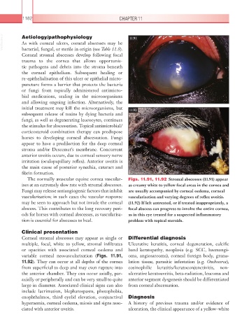
The normally avascular equine cornea vascular- Figs. 11.91, 11.92 Stromal abscesses (11.91) appear
ises at an extremely slow rate with stromal abscesses. as creamy white to yellow focal areas in the cornea and
Fungi may release antiangiogenic factors that inhibit are usually accompanied by corneal oedema, corneal
vascularisation; in such cases the vascular response vascularisation and varying degrees of reflex uveitis.
may be seen to approach but not invade the corneal (11.92) If left untreated, or if treated inappropriately, a
abscess. This contributes to the long recovery peri- focal abscess can progress to involve the entire cornea,
ods for horses with corneal abscesses, as vascularisa- as in this eye treated for a suspected inflammatory
tion is essential for abscesses to heal. problem with topical steroids.
Clinical presentation
Corneal stromal abscesses may appear as single or Differential diagnosis
multiple, focal, white to yellow, stromal infiltrates Ulcerative keratitis, corneal degeneration, calcific
or opacities with associated corneal oedema and band keratopathy, neoplasia (e.g. SCC, haemangi-
variable corneal neovascularisation (Figs. 11.91, oma, angiosarcoma), corneal foreign body, granu-
11.92). They can occur at all depths of the cornea lation tissue, parasitic infestation (e.g. Onchocerca),
from superficial to deep and may even rupture into eosinophilic keratitis/keratoconjunctivitis, non-
the anterior chamber. They can occur axially, par- ulcerative keratouveitis, beta-radiation, leucoma and
axially or peripherally and can be very small to quite anterior segment dysgenesis should be differentiated
large in diameter. Associated clinical signs can also from corneal abscessation.
include lacrimation, blepharospasm, photophobia,
enophthalmos, third eyelid elevation, conjunctival Diagnosis
hyperaemia, corneal oedema, miosis and signs asso- A history of previous trauma and/or evidence of
ciated with anterior uveitis. ulceration, the clinical appearance of a yellow–white