Page 1212 - Equine Clinical Medicine, Surgery and Reproduction, 2nd Edition
P. 1212
Eyes                                          1187


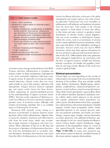
  VetBooks.ir  Table 11.9  Main causes of uveitis        traction by fibrous adhesions, or because of the globe
                                                         enlargement and zonule rupture seen with second-
                                                         ary glaucoma. Vitreal haze can occur secondary to
              • Trauma – blunt or penetrating
              • Iatrogenic from surgical trauma (i.e. intraocular surgery)  inflammatory cell infiltrate and exudation of protein
              • Systemic infections                      from the uveal tract. This exudate in the vitreous
                • Bacteria: Leptospira spp., Brucella abortus, B. melitensis,   may organise to form bands of tissue that adhere
               Borrelia burgdorferi, Salmonella spp., Streptococcus   to the retina and may contract to produce retinal
               equi, Escherichia coli, Rhodococcus (Corynebacterium)
               equi, Actinobacillus equuli, leishmaniasis, tuberculosis  detachment. In chronic uveitis, corneal degenera-
                • Parasites: Onchocerca cervicalis, Halicephalobus deletrix,   tion can occur secondary to pathological changes
               intestinal strongyles, Dirofilaria immitis  within the cornea, such as accumulation of calcium
                • Viruses: equine arteritis virus, equine infectious anaemia   in the corneal epithelial basement membrane, which
               virus, equine herpesvirus types 1 and 4, equine influenza
               virus                                     may cause disruption of the epithelium resulting in
                • Protozoal: Toxoplasma gondii, Neospora spp.  ulceration. Anterior uveitis may also lead to PIFM
                • Fungal: Cryptococcus                   formation, which may limit aqueous absorption by
              • Keratitis-associated axonal reflex uveitis  the iris and lead to physical and functional obstruc-
              • Immune-mediated/idiopathic, including equine recurrent uveitis  tion of the ICA. Other lesions that cause glaucoma
              • Neoplasia
              • Lens-induced uveitis                     with subsequent buphthalmos by interfering with
                                                         the flow of aqueous humour include lens luxation,
                                                         anterior synechiae, iris bombé and pupillary seclu-
                                                         sion. In end-stage uveitis, fibrosis of the uveal tract
          of uveitis is tissue damage and breakdown of the BAB.   results in phthisis bulbi.
          Trauma, infection, inflammation or neoplasia can
          initiate uveitis via these mechanisms. Leptospirosis  Clinical presentation
          is the most commonly implicated  infectious cause   Clinical signs can vary depending on the severity of
          of equine uveitis. In naturally occurring and experi-  inflammation, the area(s) of the uvea involved and the
          mental infections, clinical uveitis does not occur   duration of the problem. They may include variable
          until months or years after primary infection with   vision  deficits,  lacrimation,  blepharospasm,  photo-
          leptospirosis. Antigen mimicry between  Leptospira   phobia,  enophthalmos, conjunctival/episcleral  con-
          spp.  and equine ocular tissues has been demon-  gestion, corneal oedema, corneal  neovascularisation/
          strated,  supporting  an  immune-mediated  process   ciliary flush, keratic precipitates, aqueous flare, fibrin
          in the pathophysiology of equine recurrent uveitis   in the anterior chamber, hypopyon, hyphaema, mio-
          (ERU). Aberrant ocular migration of Onchocerca cer-  sis, cellular debris and/or pigment on the anterior
          vicalis microfilariae is the most commonly implicated   lens capsule, oedematous and/or hyperaemic iris,
          parasitic  cause  of  recurrent  uveitis,  although  with   yellow–green discolouration/vitreal haze, peripapil-
          modern de-worming schedules this is an unlikely   lary chorioretinitis and retinal detachment. Possible
          source of inflammation for uveitis.            sequelae include band keratopathy (white chalky
            Decreased IOP typically occurs owing to a    spiculated opacities in the cornea),  dyscoria, ante-
          decrease in production of aqueous humour result-  rior and posterior synechiae, iris bombé, iris colour
          ing from ciliary body inflammation. Aqueous flare   change, corpora nigra atrophy, cataract formation,
          is caused by exudation of protein and inflammatory   secondary glaucoma, lens subluxation/luxation, pig-
          cells from the uveal tissue,  which may  accumu-  ment changes in the iris, vitreal liquefaction/floaters,
          late in large amounts as  hypopyon  and cause syn-  vitreal traction bands, retinal detachment, depig-
          echiae. These adhesions of the iris to the lens and   mentation of the peripapillary region in a focal or
          the presence of products of inflammation that inter-  alar pattern/butterfly lesions and phthisis bulbi
          fere  with  lens  metabolism  lead  to  secondary  cata-  (Figs. 11.100–103). In many cases, posterior seg-
          ract formation. Secondary lens luxation may result   ment lesions cannot be directly visualised because of
          from the effect of inflammation on the lens zonules,   the severe anterior segment abnormalities.

