
Page 1210 - Equine Clinical Medicine, Surgery and Reproduction, 2nd Edition
P. 1210
Eyes 1185
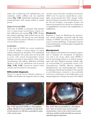
VetBooks.ir surface due to thickening of the epithelial layer, and out other causes of keratitis, including viral infection
(EHV-2 and -5), mycoses, corneal abscess, eosino-
sometimes, cellular infiltrate into the superficial
stroma (Fig. 11.96). Superficially branching corneal
neovascularisation with corneal oedema is usually philic keratoconjunctivitis, KCS, foreign bodies,
physical irritation associated with distichiasis, ecto-
present. pic cilia and entropion, neurotrophic keratitis, and
exposure keratopathy due to facial nerve dysfunction
Deep/mid-stromal IMMK or lagophthalmos associated with exophthalmia or
This form of IMMK is associated with extensive buphthalmia.
mid- to deep stromal vascularisation, oedema, cel-
lular infiltration and scarring (Fig. 11.97). Despite Diagnosis
the extensive pathology these patients may be rela- Diagnosis is based on identifying the character-
tively comfortable. The disease may cycle through istic corneal pathology associated with the three
relatively quiescent periods followed by recurrence subtypes of IMMK and ruling out other potential
of active disease episodes. causes of corneal pathology. A subsequent response
to anti-inflammatory treatment helps strengthen the
Endotheliitis diagnosis of IMMK.
In this form of IMMK the corneal endothelium
is affected, leading to varying degrees of corneal Management
oedema subsequent to disruption of the endothelial Early diagnosis and initiation of treatment is impor-
pump mechanism. The oedema may vary from mild tant to minimise the recruitment of inflamma-
to severe, with corneal decompensation (corneal tory cells to the cornea, which causes IMMK to
hydrops) occurring in some patients. Deep corneal become increasingly refractive to medical manage-
vascularisation and cellular infiltration including ment with time. Medical treatment includes topi-
clumps of cells on the endothelium may be present cal corticosteroids (1% prednisolone acetate, 0.1%
(Figs. 11.98, 11.99). Concurrent anterior uveitis dexamethasone), NSAIDs (flunixin meglumine,
and pain may be present in some patients. phenylbutazone) and immunosuppressive therapy
(cyclosporine A [CsA] and tacrolimus). The response
Differential diagnosis to anti-inflammatory treatment is not predictable
There is no specific test for definitive diagnosis of and various combinations of anti-inflammatory and
IMMK, and therefore the diagnosis relies on ruling immunosuppressive therapy may need to be trialled.
11.96 11.97
Fig. 11.96 Superficial IMMK in a Thoroughbred Fig. 11.97 Mid-stromal IMMK in a Warmblood
gelding. Multifocal corneal epithelial and superficial mare. There is extensive deep corneal stromal
stromal opacities are affecting a geographical region vascularisation, a haze to the central ventral cornea
of the ventral cornea. with dense multifocal yellowish infiltrates and some
superficial pigment deposition.