
Page 1263 - Equine Clinical Medicine, Surgery and Reproduction, 2nd Edition
P. 1263
1238 CHAPTER 12
VetBooks.ir 12.30 12.31
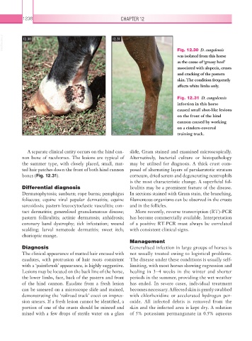
Fig. 12.30 D. congolensis
was isolated from this horse
as the cause of ‘greasy heel’
associated with alopecia, crusts
and cracking of the pastern
skin. The condition frequently
affects white limbs only.
Fig. 12.31 D. congolensis
infection in this horse
caused small shot-like lesions
on the front of the hind
cannon caused by working
on a cinders-covered
training track.
A separate clinical entity occurs on the hind can- slide, Gram stained and examined microscopically.
non bone of racehorses. The lesions are typical of Alternatively, bacterial culture or histopathology
the summer type, with closely placed, small, mat- may be utilised for diagnosis. A thick crust com-
ted hair patches down the front of both hind cannon posed of alternating layers of parakeratotic stratum
bones (Fig. 12.31). corneum, dried serum and degenerating neutrophils
is the most characteristic change. A superficial fol-
Differential diagnosis liculitis may be a prominent feature of the disease.
Dermatophytosis; sunburn; rope burns; pemphigus In sections stained with Gram stain, the branching,
foliaceus; equine viral papular dermatitis; equine filamentous organisms can be observed in the crusts
sarcoidosis; pastern leucocytoclastic vasculitis; con- and in the follicles.
tact dermatitis; generalised granulomatous disease; More recently, reverse transcription (RT)-PCR
pastern folliculitis; actinic dermatosis; anhidrosis; has become commercially available. Interpretation
coronary band dystrophy; tick infestation; wound of a positive RT-PCR must always be correlated
scalding; larval nematode dermatitis; sweet itch; with consistent clinical signs.
chorioptic mange.
Management
Diagnosis Generalised infection in large groups of horses is
The clinical appearance of matted hair encased with not usually treated owing to logistical problems.
exudates, with protrusion of hair roots consistent The disease under these conditions is usually self-
with a ‘paintbrush’ appearance, is highly suggestive. limiting, with most horses showing regression and
Lesions may be located on the back line of the horse, healing in 3–4 weeks in the winter and shorter
the lower limbs, face, back of the pastern and front periods in the summer, providing the wet weather
of the hind cannon. Exudate from a fresh lesion has ended. In severe cases, individual treatment
can be smeared on a microscope slide and stained, becomes necessary. Affected skin is gently swabbed
demonstrating the ‘railroad track’ cocci on impres- with chlorhexidine or accelerated hydrogen per-
sion smears. If a fresh lesion cannot be identified, a oxide. All infected debris is removed from the
portion of one of the crusts should be minced and skin and the infected area is kept dry. A solution
mixed with a few drops of sterile water on a glass of 5% potassium permanganate in 0.5% aqueous