
Page 1267 - Equine Clinical Medicine, Surgery and Reproduction, 2nd Edition
P. 1267
1242 CHAPTER 12
VetBooks.ir a few drops of a vitamin B solution is added to the and surrounding hair should be scrubbed for 1–2
minutes daily for 7–10 days with one of the follow-
surface of the culture medium to minimise false-
negative results. A white powdery colony is noted
or ointments containing miconazole or climbazole
within 7–28 days and long cylindrical macrospores ing treatments: 2.0% lime sulphur in water; sprays
may be detected on cytology of the growth. combined with chlorhexidine; accelerated hydrogen
Skin and hair samples may also be submitted for peroxide spray; creams containing terbinafine; or a
PCR analysis for a rapid diagnosis and speciation 0.2% enilconazole solution applied daily to twice
depending on local laboratory availability. Cutaneous weekly depending on the extent and severity of
biopsies will detect endothrix and mural folliculitis in infection. All tack should be fumigated to prevent
80% of infected cases. Trichophyton species may also spread. Some countries have access to a Trichophyton
cause acantholysis, mimicking pemphigus on histo- vaccine that reportedly has good efficacy.
pathology. For this reason, it is recommended to use
fungal stains (e.g. periodic acid–Schiff [PAS]) in any Prognosis
case of suspected pemphigus to rule out dermatophy- The prognosis is good for individuals, but hygiene
tosis as a primary cause because treatment choices are and quarantine must be strict to prevent spread.
polar opposites (i.e. immunosuppression is contrain- Infected horses should be handled last and strict
dicated in the treatment of dermatophytosis). post-contact hygiene should be in place.
Management RINGWORM CAUSED BY MICROSPORUM
Most horses develop resistance to dermatophytes
and infection is usually self-limiting. Control is Definition/overview
achieved by preventing spread between horses when Microsporosis is a common fungal infection of the
groups are involved. All crusts and infected hairs hair follicles caused by Microsporum spp. It is most
should be carefully removed and burnt. All lesions commonly caused by M. gypseum or M. canis, and
occasionally by M. equinum.
12.36 Aetiology/pathophysiology
Microsporosis is usually spread by contact with a
contaminated area (e.g. horse transport, tack, soil)
and can also be spread by biting insects and skin abra-
sion. M. gypseum is a soil saprophyte, while M. canis is
typically transmitted by asymptomatic carrier barn
cats and M. equinum is spread from infected horses,
their tack or blankets. Unlike Trichophyton spp.,
which invade the hair shaft resulting in endothrix
infection, Microsporum spp. microspores invade the
outer hair shaft, causing ectothrix infection, visu-
alised by direct examination of infected hair shafts
or via histopathology and PAS staining. In either
case, the infection fails to destroy all the hair in
the infected area so that a clean pluck rarely occurs,
leaving a moth-eaten look to the hair coat.
Clinical presentation
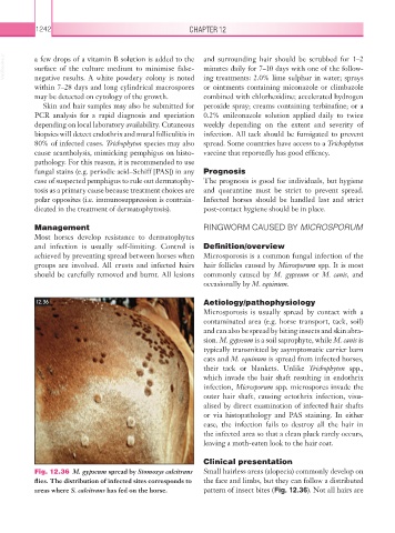
Fig. 12.36 M. gypseum spread by Stomoxys calcitrans Small hairless areas (alopecia) commonly develop on
flies. The distribution of infected sites corresponds to the face and limbs, but they can follow a distributed
areas where S. calcitrans has fed on the horse. pattern of insect bites (Fig. 12.36). Not all hairs are