
Page 1266 - Equine Clinical Medicine, Surgery and Reproduction, 2nd Edition
P. 1266
Skin 1241
VetBooks.ir INFECTIOUS CAUSES – FUNGAL or foal (e.g. the girth). Actively infected sites are pru-
(DERMATOPHYTOSIS)
ritic. Initially single lesions occur, then the infected
RINGWORM CAUSED BY TRICHOPHYTON area becomes covered with multiple, often coalesc-
ing lesions (Fig. 12.35). The hair plucks easily 10–15
days post infection. Trichophyton may also result in a
Definition/overview crusting dermatitis similar in appearance to pemphi-
This is a common fungal infection of the hair fol- gus foliaceous, caused by the organism’s acantholytic
licles caused by Trichophyton spp. T. equinum is the effects due to the release of an exfoliative toxin.
most common cause of dermatophytosis in horses.
It is a highly contagious disease affecting horses of Differential diagnosis
all ages. It is spread by contact with a source of con- Dermatophilosis; Microsporum gypseum infection;
taminated material or from horse to horse. sweet itch; insect hypersensitivity; mite infestation;
equine sarcoidosis; equine granulomatous enteri-
Aetiology/pathophysiology tis; alopecia areata; anhidrosis; actinic dermatitis;
Most common are T. equinum var. equinum, T. equi- wound scalding; Malassezia spp. infection.
num var. autotrophicum and, less commonly, T. ver-
rucosum, T. bullosum and T. mentagrophytes. The Diagnosis
organisms are found on horse hair or fomites and Hairs plucked from a fresh lesion are cleaned with
rarely in soil. Infection occurs more readily if the chlorlactophenol, then examined on a clear slide
skin is abraded (e.g. rubbed by grooming brush, with warm 30% potassium hydroxide. Hyphae and
girth) and contaminated with the organism. The large endothrix spores will be seen. Trichophyton-
surface of the hair shaft is invaded by arthrospores, infected hairs do not fluoresce under black UV light.
which migrate down to invade the hair follicle. The hair is cultured on Sabouraud’s agar or rapid
Clinical disease appears 9–15 days post exposure. sporulating medium (RSM) and dermatophyte test
medium (DTM) at 25°C (77°F). DTM is essentially
Clinical presentation Sabouraud’s dextrose agar containing cyclohexi-
Loss of hair and scaling of skin occur. In the early mide, gentamicin and chlortetracycline as antifun-
stages, hair follicles become erect in a circular area gal and antibacterial agents and to which the pH
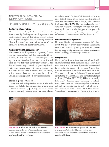
5–20 mm in diameter (Fig. 12.34). Lesions can occur indicator phenol red has been added. Also, because
wherever contaminated equipment contacts the horse Trichophyton is dependent on thiamine for growth,
12.34 12.35
Fig. 12.34 Early infection with T. equinum var. Fig. 12.35 An older dermatophyte infection with
equinum due to the use of a contaminated girth a large area of alopecia. The early lesions have
14 days earlier is seen as small areas of alopecia and coalesced, with a secondary infected area of smaller
small papules with erect hairs. peripheral lesions.