
Page 1286 - Equine Clinical Medicine, Surgery and Reproduction, 2nd Edition
P. 1286
Skin 1261
VetBooks.ir head, neck and limbs and very rarely in the parageni- 12.55
tal region. Geographical variation may potentially be
associated with feeding patterns of different flies and
hence transmission of the virus to different sites. The
fly vector transmission hypothesis is also supported by
the lack of equine sarcoids in Norway, a country with-
out biting insects. A study of sarcoid cases in the UK
showed single and small numbers of sarcoids (2–8 sar-
coids per horse) to be uncommon, whereas 14–84% of
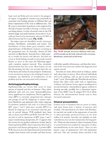
affected horses had 10 or more (Fig. 12.55).
Six major types are described: occult, verrucous,
nodular, fibroblastic, malevolent and mixed. The
distribution of these shows great variation, with a
preponderance of fibroblastic tumours occurring in
the paragenital area. In Australia, almost all limb Fig. 12.55 Sarcoid. An 8-year-old horse with a mix
sarcoids are fibroblastic. Sarcoids have a high capac- of (35) sarcoids on the body and neck with verrucous,
ity to invade the dermis and subcutis. Sarcoids can fibroblastic, mixed and occult forms.
occur in fresh healing wounds in previously normal
horses, or recur at the same site following appar-
ently complete surgical removal. True metastatic with other spindle cell tumours, and therefore detec-
dissemination does not occur. Most lesions are not tion of the virus does not confirm the diagnosis of an
life-threatening, but they can severely limit the use equine sarcoid.
of the horse and reduce its sale prospects. Euthanasia Sarcoids are more often noted in donkeys, mules
is not uncommon owing to the prolonged nature of and zebras than in horses. Most affected individuals
treatment, the likelihood of recrudescence of the tend to be geldings, with an age at onset between
problem and the cost associated with treatment. 1 and 7 years. Thoroughbreds, Warmbloods and those
horses that often work cattle such as Appaloosas,
Aetiology/pathophysiology Arabians and Quarter horses appear predisposed to
Papillomaviridae are viruses that occur in many sarcoid formation. Standardbreds appear unlikely to
species of animals and also in humans. They infect develop sarcoids, possibly due to decreased expres-
epithelial cells and can cause hyperproliferation, sion of the MHC class II antigen W13 ELA allele,
warts, papillomas or condylomas. Bovine papilloma- whereas the aforementioned breeds tend to have
virus (BPV) is currently categorised into subtypes increased expression.
and groups (A and B). Subgroup A viruses trans-
form fibroblasts and epithelial cells, while subgroup Clinical presentation
B transform epithelial cells only. It is believed that Lesions occur in locations that are prone to injury,
BPV types 1 and 2 (and more recently BPV13 in and some horses have a history of previous skin
Brazil) of subgroup A are associated with the gen- wounds. The introduction of a horse with fibroblas-
esis of sarcoid disease. PCR methods have been tic sarcoids can result in the appearance of sarcoids
able to detect viral DNA and RNA from 88–91% of in other previously uninfected horses on the farm
sarcoids as well as expression of BPV types 1 and 2 within 6–8 months. Sarcoids can multiply on indi-
major transforming oncoprotein, E5. It appears that vidual horses, but can also remain static for long peri-
the viruses do not produce infectious virions, but by ods. There are also instances where treatment of one
downregulating MHC class I expression they lead to or several lesions has resulted in regression of other
persistence and disease pathogenesis. A positive BPV untreated lesions on the same horse. There are also
PCR, however, has also been detected in normal skin known cases of spontaneous full and permanent
samples from horses with sarcoids and from horses self-cure. The one certainty about sarcoids is the