
Page 1284 - Equine Clinical Medicine, Surgery and Reproduction, 2nd Edition
P. 1284
Skin 1259
VetBooks.ir 12.53 Prognosis
New cases show a better response than chronic cases.
Calcified lesions need surgical removal.
EQUINE UNILATERAL
PAPULAR DERMATOSIS
Definition/overview
This condition is most frequently seen in yearlings
and 2-year-old horses. Papules and nodules appear in
multiples, usually forming a loose group in a circular
arrangement.
Fig. 12.53 Equine eosinophilic granuloma. One large Aetiology/pathophysiology
calcified nodule and several small hard subcutaneous The aetiology is unknown. It is most commonly
nodules are present on the side of the chest. reported in Quarter horses.
Clinical presentation
Differential diagnosis Numerous, even sized, firm, round, well-circum-
Mastocytoma; epidermoid and dermoid cysts; insect scribed nodules are evident. There is no alopecia
bites; unilateral papular dermatosis; hypodermiasis; or pruritus and the nodules are non-ulcerative
amyloidosis; phaeohyphomycosis; panniculitis. and non-painful (Fig. 12.54). Fresh nodules may
occur in almost concentric rings around the origi-
Diagnosis nal lesions.
Biopsy and histopathology, which show foci of
degeneration of collagen hairs associated with heavy Differential diagnosis
eosinophilic infiltration, are the best diagnostic Dermatophytosis (before hair is lost); Stomoxys calci-
techniques. trans bites; sweet itch and other insect bites; oncho-
cercal filariasis.
Management
Glucocorticoids are the principal means of treat-
ing these lesions. If a single lesion is noted, 12.54
intralesional or sublesional injection of 5 mg tri-
amcinolone acetonide every 2 weeks for three
treatments provides a non-surgical option. If an
incomplete resolution is noted with this protocol,
or there is concern regarding laminitis and other
adverse effects associated with the use of gluco-
corticoids, surgical extirpation or CO or diode
2
laser ablation should be considered, especially if
lesions have become calcified. When multiple
lesions are present, prednisolone (1–2 mg/kg/day
7–10 days, then tapering completely off medica-
tion within 3–4 weeks) is likely to help with this
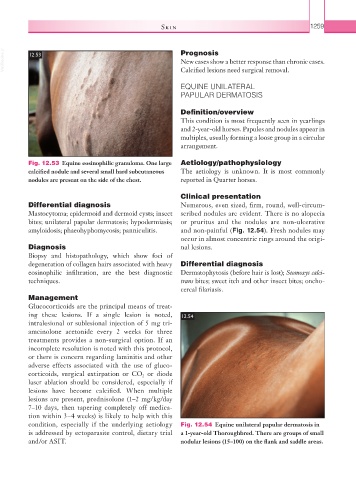
condition, especially if the underlying aetiology Fig. 12.54 Equine unilateral papular dermatosis in
is addressed by ectoparasite control, dietary trial a 1-year-old Thoroughbred. There are groups of small
and/or ASIT. nodular lesions (15–100) on the flank and saddle areas.