
Page 1282 - Equine Clinical Medicine, Surgery and Reproduction, 2nd Edition
P. 1282
Skin 1257
VetBooks.ir IMMUNE-MEDIATED NODULES OR Aetiology/pathophysiology
SWELLINGS ASSOCIATED WITH
These are congenital, developmental and possi-
CONGENITAL, DEVELOPMENTAL
AND POSSIBLY HEREDITARY bly hereditary lesions. They are thought to be due
to embryonal displacement of ectoderm into the
DYSFUNCTIONS subcutis.
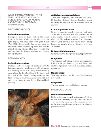
DENTIGEROUS CYSTS Clinical presentation
Single to multiple nodules, covered with skin,
Definition/overview 10–15 mm in diameter and usually found on the
Dentigerous cysts are hard swellings that occur dorsal midline from the withers to rump will be
between the base of the ear and the eye (tem- observed. The nodules contain soft keratinaceous
poral region) and arise from tooth germ tissue caseous grey material and coiled hairs. Young
(Fig. 12.52). Occasionally, they are also found on horses are usually affected, between birth and
the cranial vault or maxillary sinus and contain 18 months.
enamel-forming tissue. They may contain one
tooth or more. Drainage may occur near the ear Differential diagnosis
through the skin. Epidermoid cysts; dentigerous cysts; hypodermiasis.
DERMOID CYSTS Diagnosis
The location and clinical picture are suggestive.
Definition/overview Excisional biopsy shows a cyst wall lined with
Dermoid cysts are single or multiple, firm to stratified squamous epithelium containing adnexal
fluctuant, smooth, round cysts, usually with nor- structures.
mal overlying haired skin. They most commonly
occur along the dorsal midline of the thorax and Management
back, and while a breed predisposition has not Total surgical ablation of the cyst wall and contents
been proven, an Australian survey indicated that is required.
they occur more frequently in Thoroughbred
yearlings. Prognosis
The prognosis is very good following surgery.
12.52 EPIDERMOID CYSTS
Definition/overview
Epidermoid cysts are usually solitary cysts, 7–30 mm
in diameter, occurring on the head and limbs.
They are freely movable, well-circumscribed, firm
to fluctuant on palpation and contain a yellow to
grey mucoid fluid.
Fig. 12.52 Dentigerous cyst at the base of the ear.