
Page 1278 - Equine Clinical Medicine, Surgery and Reproduction, 2nd Edition
P. 1278
Skin 1253
VetBooks.ir 12.48 12.49
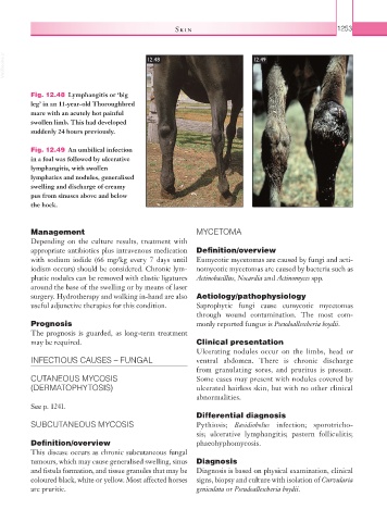
Fig. 12.48 Lymphangitis or ‘big
leg’ in an 11-year-old Thoroughbred
mare with an acutely hot painful
swollen limb. This had developed
suddenly 24 hours previously.
Fig. 12.49 An umbilical infection
in a foal was followed by ulcerative
lymphangitis, with swollen
lymphatics and nodules, generalised
swelling and discharge of creamy
pus from sinuses above and below
the hock.
Management MYCETOMA
Depending on the culture results, treatment with
appropriate antibiotics plus intravenous medication Definition/overview
with sodium iodide (66 mg/kg every 7 days until Eumycotic mycetomas are caused by fungi and acti-
iodism occurs) should be considered. Chronic lym- nomycotic mycetomas are caused by bacteria such as
phatic nodules can be removed with elastic ligatures Actinobacillus, Nocardia and Actinomyces spp.
around the base of the swelling or by means of laser
surgery. Hydrotherapy and walking in-hand are also Aetiology/pathophysiology
useful adjunctive therapies for this condition. Saprophytic fungi cause eumycotic mycetomas
through wound contamination. The most com-
Prognosis monly reported fungus is Pseudoallescheria boydii.
The prognosis is guarded, as long-term treatment
may be required. Clinical presentation
Ulcerating nodules occur on the limbs, head or
INFECTIOUS CAUSES – FUNGAL ventral abdomen. There is chronic discharge
from granulating sores, and pruritus is present.
CUTANEOUS MYCOSIS Some cases may present with nodules covered by
(DERMATOPHYTOSIS) ulcerated hairless skin, but with no other clinical
abnormalities.
See p. 1241.
Differential diagnosis
SUBCUTANEOUS MYCOSIS Pythiosis; Basidiobolus infection; sporotricho-
sis; ulcerative lymphangitis; pastern folliculitis;
Definition/overview phaeohyphomycosis.
This disease occurs as chronic subcutaneous fungal
tumours, which may cause generalised swelling, sinus Diagnosis
and fistula formation, and tissue granules that may be Diagnosis is based on physical examination, clinical
coloured black, white or yellow. Most affected horses signs, biopsy and culture with isolation of Curvularia
are pruritic. geniculata or Pseudoallescheria boydii.