
Page 1279 - Equine Clinical Medicine, Surgery and Reproduction, 2nd Edition
P. 1279
1254 CHAPTER 12
VetBooks.ir Management Diagnosis
Diagnosis is based on identification of hyphae in
Some cases respond well to surgical removal and use
of systemic iodides. Some prove difficult and require
microscopic examination for correct identification.
repeated treatments. tissue biopsies, culture on Sabouraud’s agar and
Amplification of DNA targets can be performed for
Prognosis species identification.
Prolonged treatment may be unsuccessful.
Management
PHAEOHYPHOMYCOSIS Systemic iodide treatment is indicated. Amphotericin
B therapy is unsuccessful. Fluconazole (5 mg/kg
Definition/overview q24 h) has been suggested. Topical application of eti-
This is a chronic subcutaneous and systemic fungal sazole in DMSO has been reported.
disease, often presenting as small multiple subcutane-
ous nodules. Drechslera spicifera has previously been Prognosis
isolated. A guarded prognosis should be given.
Aetiology/pathophysiology PYTHIOSIS (PHYCOMYCOSIS)
Phaeohyphomycosis is caused by various saprophytic
soil fungi contaminating wounds. See p. 1213.
Clinical presentation SPOROTRICHOSIS
Small, black, denuded plaques containing papules
and pustules will be identified. Multiple small, Definition/overview
fibrotic, subcutaneous nodules can occur on the Sporotrichosis is a cutaneous and subcutaneous nodular
neck, body and limbs. intermediate fungal skin infection that extends to the
lymphatics, resulting in ulcerative lymphatic cording.
Differential diagnosis
Eosinophilic granuloma; infectious granuloma; neo- Aetiology/pathophysiology
plasia; foreign body granuloma. Sporotrichosis is a zoonotic mycosis caused by a
dimorphic fungus, Sporothrix schenckii, that occurs as
a yeast form in tissues and a telomorph form in soil
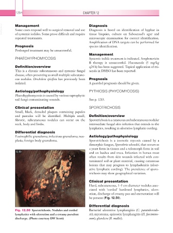
12.50 and on bushes and trees. Infection in horses most
often results from skin wounds infected with con-
taminated soil or plant material, causing cutaneous
lesions that may progress to lymphadenitis (ulcer-
ative lymphatic cording). The prevalence of sporo-
trichosis may show geographical variation.
Clinical presentation
Hard, subcutaneous, 1–5 cm diameter nodules asso-
ciated with ‘corded’ hardened lymphatics, ulcer-
ation, discharge of creamy pus and encrustation will
be present (Fig. 12.50).
Differential diagnosis
Fig. 12.50 Sporotrichosis. Nodules and corded Bacterial ulcerative lymphangitis (C. paratuberculo-
lymphatics with ulceration and a creamy purulent sis); mycetoma; epizootic lymphangitis (H. farcimino-
discharge. (Photo courtesy DW Scott) sum); glanders (B. mallei).