
Page 1295 - Equine Clinical Medicine, Surgery and Reproduction, 2nd Edition
P. 1295
1270 CHAPTER 12
VetBooks.ir Diagnosis 12.67
Diagnosis is based on immunohistological confir-
mation that the nodules are composed of densely
packed lymphoblastic cells expressing CD79a, and
numerous small, round, CD3-positive T lympho-
cytes. Abnormal circulating lymphocytes are seen in
25–50% of cases. Decreased IgM level is often seen
in horses with lymphoma; however, the sensitivity is
poor (~28%) while specificity is fairly good (~88%).
Management
Wide surgical removal is indicated. Other than proges-
tins and surgical removal of ovarian tumours, therapy
may involve identifying and eliminating the antigenic
stimulus, considering antiviral therapy, or predniso-
lone (1–2 mg/kg q24 h with taper) and/or lomustine
chemotherapy (60–90 mg/m every 30 days).
2
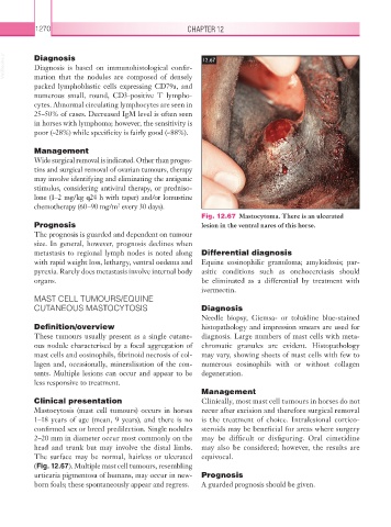
Fig. 12.67 Mastocytoma. There is an ulcerated
Prognosis lesion in the ventral nares of this horse.
The prognosis is guarded and dependent on tumour
size. In general, however, prognosis declines when
metastasis to regional lymph nodes is noted along Differential diagnosis
with rapid weight loss, lethargy, ventral oedema and Equine eosinophilic granuloma; amyloidosis; par-
pyrexia. Rarely does metastasis involve internal body asitic conditions such as onchocerciasis should
organs. be eliminated as a differential by treatment with
ivermectin.
MAST CELL TUMOURS/EQUINE
CUTANEOUS MASTOCYTOSIS Diagnosis
Needle biopsy, Giemsa- or toluidine blue-stained
Definition/overview histopathology and impression smears are used for
These tumours usually present as a single cutane- diagnosis. Large numbers of mast cells with meta-
ous nodule characterised by a focal aggregation of chromatic granules are evident. Histopathology
mast cells and eosinophils, fibrinoid necrosis of col- may vary, showing sheets of mast cells with few to
lagen and, occasionally, mineralisation of the con- numerous eosinophils with or without collagen
tents. Multiple lesions can occur and appear to be degeneration.
less responsive to treatment.
Management
Clinical presentation Clinically, most mast cell tumours in horses do not
Mastocytosis (mast cell tumours) occurs in horses recur after excision and therefore surgical removal
1–18 years of age (mean, 9 years), and there is no is the treatment of choice. Intralesional cortico-
confirmed sex or breed predilection. Single nodules steroids may be beneficial for areas where surgery
2–20 mm in diameter occur most commonly on the may be difficult or disfiguring. Oral cimetidine
head and trunk but may involve the distal limbs. may also be considered; however, the results are
The surface may be normal, hairless or ulcerated equivocal.
(Fig. 12.67). Multiple mast cell tumours, resembling
urticaria pigmentosa of humans, may occur in new- Prognosis
born foals; these spontaneously appear and regress. A guarded prognosis should be given.