
Page 1298 - Equine Clinical Medicine, Surgery and Reproduction, 2nd Edition
P. 1298
Skin 1273
VetBooks.ir Management 12.71
Complete surgical removal is required. Tumours
recur at a different site in the eyelid in approximately
25–50% of all operated cases. Injection of tumours
with BCG (1–4 injections, 28 days apart) has shown
some success.
Prognosis
The prognosis is guarded. The tumours are persis-
tent, but multiple surgeries will prolong useful life.
SQUAMOUS CELL CARCINOMA Fig. 12.71 Squamous cell carcinoma of the prepuce.
This depigmented skin tumour was moveable with
Definition/overview preputial skin and was completely removed surgically.
SCC is the second most common tumour diagnosed
in horses and accounts for 20% of equine neoplasms. 12.72
Breeds more commonly affected include draught
breeds, Appaloosas, American Paints and Pintos.
Aetiology/pathophysiology
SCC is a malignant tumour arising from keratino-
cytes. Exposure of the skin to actinic (solar) radiation
due to high altitude, decreased skin pigmentation
and sparse hair coat is frequently involved. The irri-
tant nature of equine smegma is implicated in male
genital SCC (Fig. 12.71). Irritation due to these Fig. 12.72 Squamous cell carcinoma at the
factors may be a trigger for accelerated growth. mucocutaneous junction of the nose.
Clinical presentation 12.73
SCC is characterised by a small granulating sore
that may be depressed below skin level, erod-
ing into normal tissue. There is often malodour
even with early lesions. It commonly affects the
non-pigmented mucocutaneous junctions includ-
ing the ocular conjunctiva and the external geni-
talia but can also involve other areas of the body
including the perioral and perinasal regions, the
aural canal and perianal tissue. On the penis and
prepuce they may vary from cauliflower-like to
erosive. On the vulva and anus they are often slow
growing. On the eye and eyelid they often start as
white, raised plaques at the edge of the lid or cor-
neal/scleral junction, and may progress rapidly to
granulomatous and ulcerated lesions. Around the
nose and mouth they appear as a depressed ulcer
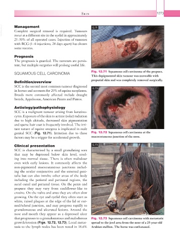
that progresses to a granulomatous and malodorous Fig. 12.73 Squamous cell carcinoma with metastatic
growth/erosion (Figs. 12.72, 12.73). Local metas- spread to the jowl area from the nose of a 25-year-old
tasis to the lymph nodes has been noted in 18.6% Arabian stallion. The horse was euthanased.