
Page 1303 - Equine Clinical Medicine, Surgery and Reproduction, 2nd Edition
P. 1303
1278 CHAPTER 12
VetBooks.ir Differential diagnosis Janus–kinase inhibitors in humans has shown great
promise and may be a consideration for horses.
Dermatophytosis; dermatophilosis; occult sarcoid;
anagen effluvium (defluxion); telogen effluvium
of clinical signs has been documented.
(defluxion); follicular dysplasia. Lastly, benign neglect with spontaneous resolution
Diagnosis Prognosis
Non-specific alopecia is present, with mane and The likelihood of complete and permanent remis-
tail dystrophy. Skin biopsies of multiple stages of sion is poor, but the condition is purely cosmetic.
alopecia will help to confirm the diagnosis, espe-
cially in the early stages. Often serial step-sections EQUINE LINEAR KERATOSIS
are required to find characteristic lesions of bulbi- (EQUINE LINEAR ALOPECIA)
tis. Microscopically, the hair bulbs, inner and outer
root sheaths of inferior segments and perifollicular Definition/overview
regions are infiltrated by small to moderate numbers This is an idiopathic, possibly inherited, dermatosis
of small lymphocytes characterised as a ‘swarm of characterised by unilateral, linear, vertical bands of
bees’. Similar inflammation is occasionally evident hyperkeratosis on the neck, thorax and upper hindlimbs
in isthmus follicular walls as well as some apo- (Fig. 12.81). Linear keratosis is more likely to be a
crine glands. Immunohistochemistry confirms that keratinisation disorder and not a classically defined
the small lymphocytes are CD3+ T lymphocytes. immune-mediated condition; however, because lym-
Diagnostic histopathological evidence may not be phocytic mural inflammation is sometimes noted on
present in more chronic lesions. histopathology it has been included in this section.
Management Aetiology/pathophysiology
Topical tacrolimus 0.1%, systemic, topical or intra- Equine linear keratosis is an uncommon to rare, pos-
lesional corticosteroids and minoxidil have been sibly inherited, dermatosis of unclear pathogenesis.
attempted with variable success. Recently, use of It resembles a linear epidermal hamartoma.
12.80 12.81
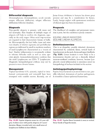
Fig. 12.80 Equine alopecia areata in a 13-year-old Fig. 12.81 Equine linear keratosis is seen as vertical
grey Arabian mare with patchy alopecia of 5 years’ linear bands of hyperkeratosis.
duration. Biopsy revealed only very small areas of
lymphocytic bulbitis in hair follicles.