
Page 1304 - Equine Clinical Medicine, Surgery and Reproduction, 2nd Edition
P. 1304
Skin 1279
VetBooks.ir Clinical presentation EQUINE PEMPHIGUS FOLIACEUS
The condition occurs at between 1 and 5 years of age
and is seen most commonly in Quarter horses. One Definition/overview
or more vertically arranged bands of alopecia and Pemphigus foliaceus is an autoimmune disease char-
hyperkeratotic papules and plaques associated with acterised by an exfoliative dermatitis. It is a pus-
scaling/crusting are present. There is no pruritus tular disease with the formation of heavy crusts
or pain. (Figs. 12.82, 12.83). Appaloosas may be predisposed.
Differential diagnosis Aetiology/pathophysiology
Chronic hyperkeratosis from irritant drugs; Pemphigus foliaceus is caused by an autoimmune
dermatophilosis. attack targeting the desmosomes in the upper lay-
ers of the epidermis. Destruction of the desmo-
Diagnosis somes causes immature keratinocytes to exfoliate
Biopsy should be performed for histopathology. The prematurely and form subcorneal pustules. These
primary change is a lymphocytic mural folliculitis. pustules are frequently ruptured, leaving epidermal
collarettes, crusts and scale as the clinical lesions.
Management Rarely, pemphigus can occur secondary to preg-
Use of a keratolytic agent, such as 5% salicylic acid nancy, foaling, drugs and vaccinations.
ointment or antiseborrhoeic shampoo, reduces the
amount of hyperkeratosis. Topical tacrolimus 0.1% Clinical presentation
and topical retin-A have been used with some suc- Early cases show transient vesicles, erosion, epider-
cess. Remission ceases when treatment stops. mal collarettes, crusting, scaling, fever, depression,
weight loss, coronary band lesions, variable pruritus
Prognosis and pain. Advanced cases show severe crusting and
These lesions are generally present for life and scaling, alopecia and poor appetite. Pitting oedema
will not completely resolve, but they are typically of the limbs and the ventral abdomen and sheath is
asymptomatic. also common.
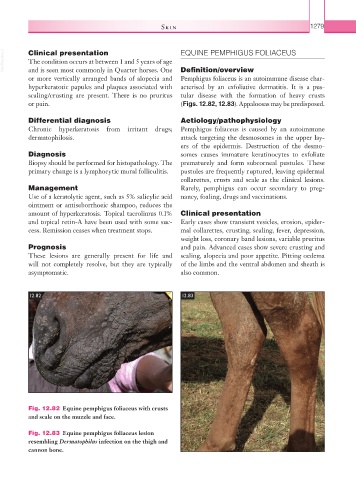
12.82 12.83
Fig. 12.82 Equine pemphigus foliaceus with crusts
and scale on the muzzle and face.
Fig. 12.83 Equine pemphigus foliaceus lesion
resembling Dermatophilus infection on the thigh and
cannon bone.