
Page 1307 - Equine Clinical Medicine, Surgery and Reproduction, 2nd Edition
P. 1307
1282 CHAPTER 12
VetBooks.ir by local contact, injection (insect allergens, the location of the oedema (e.g. whether it is in the
The lesion may present differently depending on
drugs), ingestion (chemical, feeds) or inhalation
and transepidermal absorption (pollen, dust,
chemicals, moulds). upper layers of the dermis or subcutaneous tissue):
• Physical urticaria: non-immunological • ‘Oozing’ urticaria: where dermal oedema is
pathogenesis. severe, serum may ooze from the skin surface,
• Dermatographism: wheal developing from a forming crusted lesions. Care should be taken
blunt scratch on skin. to distinguish this from an erosive/ulcerative
• Urticaria due to cold, heat or light. process, pyoderma or pemphigus foliaceus. The
• Exercise-induced urticaria. lesion must still pit on pressure.
• Angioedema (angioneurotic oedema): a
Clinical presentation subcutaneous form of urticaria that tends to be
The onset of the condition can be acute to per- more diffuse owing to lack of restraint against
acute, with signs developing within minutes up to spread in the subcutis. It usually involves the
a few hours. An oedematous lesion of the skin or head and extremities and is more indicative of
mucous membrane develops, called a wheal. This is a systemic and serious disease than urticaria.
a flat-topped papule/nodule with steep walled sides, Pruritus may or may not be present.
which pits on pressure. Some have slightly depressed
centres. Wheals vary in size and shape and may be Diagnosis
divided into: Initially, diagnosis is mostly based on clinical signs
and history. Biopsy may help to rule out other poten-
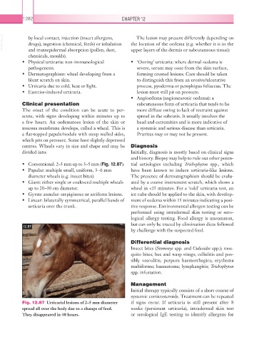
• Conventional: 2–3 mm up to 3–5 mm (Fig. 12.87). tial aetiologies including Trichophyton spp., which
• Papular: multiple small, uniform, 3–6 mm have been known to induce urticaria-like lesions.
diameter wheals (e.g. insect bites). The presence of dermatographism should be evalu-
• Giant: either single or coalesced multiple wheals ated by a coarse instrument scratch, which shows a
up to 20–30 cm diameter. wheal in <15 minutes. For a ‘cold’ urticaria test, an
• Gyrate annular: serpiginous or arciform lesions. ice cube should be applied to the skin, with develop-
• Linear: bilaterally symmetrical, parallel bands of ment of oedema within 15 minutes indicating a posi-
urticaria over the trunk. tive response. Environmental allergen testing can be
performed using intradermal skin testing or sero-
logical allergy testing. Food allergy is uncommon,
but can only be traced by elimination diets followed
12.87
by challenge with the suspected feed.
Differential diagnosis
Insect bites (Stomoxys spp. and Culicoides spp.); mos-
quito bites; bee and wasp stings; cellulitis and pos-
sibly vasculitis; purpura haemorrhagica; erythema
multiforme; haematoma; lymphangitis; Trichophyton
spp. infestation.
Management
Initial therapy typically consists of a short course of
systemic corticosteroids. Treatment can be repeated
Fig. 12.87 Urticarial lesions of 2–5 mm diameter if signs recur. If urticaria is still present after 8
spread all over the body due to a change of feed. weeks (persistent urticaria), intradermal skin test
They disappeared in 48 hours. or serological IgE testing to identify allergens for