
Page 1311 - Equine Clinical Medicine, Surgery and Reproduction, 2nd Edition
P. 1311
1286 CHAPTER 12
VetBooks.ir and production of antimelanocyte antibodies, as diagnosis. At early stages, there is mild lympho-
cytic infiltration with multifocal lymphocytic exo-
detected in humans, dogs, cats and Arabian horses.
Alternatively, autotoxicity has been proposed, result-
melanocytes.
ing from susceptibility of melanocytes to melanin cytosis. At later stages, there is complete absence of
precursors (dopachrome) or inhibition of free radi-
cal scavengers (thioredoxinreductase). Several com- Management
pounds such as phenols may exacerbate pigment loss Treatment in humans includes topical/systemic cal-
from affected melanocytes. cineurin inhibitors (e.g. tacrolimus 0.1%), vitamin D,
antioxidants and narrow band UVB light. In horses,
Clinical presentation there is no reliably effective treatment. Topically
Depigmented circular spots up to 1 cm diameter, applied or systemically delivered L-phenylalanine
which increase in number rather than size, are pres- may help to stimulate more pigment production
ent (Fig. 12.92). Occasional white patches and leuco- because this amino acid is the precursor for tyrosine,
derma depigmentation may be evident. The condition an essential component in melanogenesis. Rarely,
may wax and wane in intensity, but is usually perma- spontaneous resolution has been reported.
nent. Alopecia occasionally occurs around the eyes.
Prognosis
Differential diagnosis Partial spontaneous recovery has been reported, but
Copper deficiency; equine CLE; leucoderma; is uncommon. The number of spots may increase
Appaloosa parentage. over time. Given that this condition is likely to be
heritable, affected horses should not be used for
Diagnosis breeding.
Clinical appearance and absence of injury are sug-
gestive. Wetting the horse with water allows depig- PHOTOSENSITISATION DERMATITIS
mentation to become more obvious. History may
indicate Arabian or Welsh Mountain pony heritage. Definition/overview
Leucoderma may not be synchronised with the area This condition is caused by UV radiation and facili-
of associated leucotrichia. Skin biopsy can assist tated by lack of pigment and hair. It may be acute or
chronic.
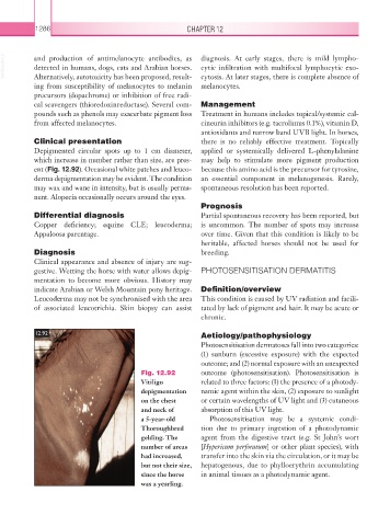
12.92 Aetiology/pathophysiology
Photosensitisation dermatoses fall into two categories:
(1) sunburn (excessive exposure) with the expected
outcome; and (2) normal exposure with an unexpected
Fig. 12.92 outcome (photosensitisation). Photosensitisation is
Vitiligo related to three factors: (1) the presence of a photody-
depigmentation namic agent within the skin, (2) exposure to sunlight
on the chest or certain wavelengths of UV light and (3) cutaneous
and neck of absorption of this UV light.
a 5-year-old Photosensitisation may be a systemic condi-
Thoroughbred tion due to primary ingestion of a photodynamic
gelding. The agent from the digestive tract (e.g. St John’s wort
number of areas [Hypericum perforatum] or other plant species), with
had increased, transfer into the skin via the circulation, or it may be
but not their size, hepatogenous, due to phylloerythrin accumulating
since the horse in animal tissues as a photodynamic agent.
was a yearling.