
Page 1316 - Equine Clinical Medicine, Surgery and Reproduction, 2nd Edition
P. 1316
Skin 1291
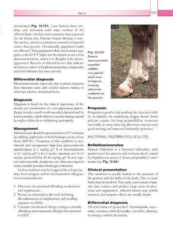
VetBooks.ir demarcated (Fig. 12.101). Later lesions show ero- 12.101
sions and ulceration with some oedema of the
affected limb, which is more extensive than expected
for the lesion size. Chronic lesions develop a wart-
like surface, which is resistant to removal and painful
rather than pruritic. Occasionally, pigmented limbs
are affected. Non-pigmented skin involvement sug- Fig. 12.101
gests a role for UV light, but the disease is not a true Pastern
photosensitisation, rather it is thought to be photo- leucocytoclastic
aggravated. Records of affected horses also indicate vasculitis
no known contact with photosensitising compounds, exhibits
and liver function has been normal.
very painful
small areas
Differential diagnosis of alopecia,
Photosensitisation, especially due to plants (requires crusting
liver function tests and careful history taking to and serum
eliminate plants); dermatophilosis.
exudation on
the pastern.
Diagnosis
Diagnosis is based on the clinical appearance of the
disease and involvement of a non-pigmented pastern. Prognosis
Biopsy reveals a small-vessel vasculitis characterised by Prognosis is good to fair pending the clinician’s abil-
leucocytoclasis, which refers to vascular damage caused ity to identify the underlying trigger factor. Some
by nuclear debris from infiltrating neutrophils. patients require life-long pentoxifylline treatment
once daily or every other day. Recovery requires very
Management good nursing and improved husbandry practices.
Affected areas should be protected from UV radiation
by stabling, application of limb bandages or use of sun BACTERIAL PASTERN FOLLICULITIS
block (SPF30+). Treatment of this condition is mul-
timodal and incorporates high-dose glucocorticoids Definition/overview
(prednisolone at 1 mg/kg q12 h or dexamethasone Pastern folliculitis is a bacterial folliculitis, with
at 0.1 mg/kg q24 h for 2 weeks, tapering over 4–12 pyoderma of the pastern and coronary band, caused
weeks), pentoxifylline (8–10 mg/kg q12 h) and topi- by Staphylococcus aureus, S. hyicus and possibly S. inter-
cal corticosteroids. Antibiotics are often not required medius (see Fig. 12.94).
unless notable purulent discharge is detected.
As this condition may be triggered by a drug reac- Clinical presentation
tion, food antigens and/or environmental allergens, The condition is usually limited to the posterior of
it is recommended to: the pastern and the bulbs of the heels. One or more
limbs may be involved. Very early cases consist of pap-
1 Eliminate the potential offending medications ules that coalesce and produce large areas of ulcer-
and supplements. ation and suppuration. Affected horses may exhibit
2 Pursue an elimination diet trial including lameness, but systemic effects are usually absent.
discontinuation of supplements and avoiding
exposure to alfalfa. Differential diagnosis
3 Consider intradermal allergy testing to identify All other forms of ‘greasy heel’: Dermatophilus, myce-
offending environmental allergies for inclusion toma, coronary band dystrophy, vasculitis, choriop-
in ASIT. tic mange, contact dermatitis.