
Page 1346 - Equine Clinical Medicine, Surgery and Reproduction, 2nd Edition
P. 1346
Wound management and infections of synovial structures 1321
VetBooks.ir 13.38A 13.38B
13.38C
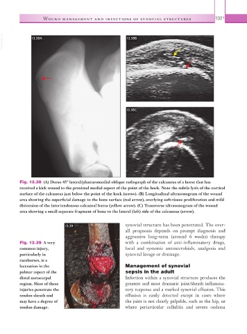
Fig. 13.38 (A) Dorso 45° lateral/plantaromedial oblique radiograph of the calcaneus of a horse that has
received a kick wound to the proximal medial aspect of the point of the hock. Note the subtle lysis of the cortical
surface of the calcaneus just below the point of the hock (arrow). (B) Longitudinal ultrasonogram of the wound
area showing the superficial damage to the bone surface (red arrow), overlying soft-tissue proliferation and mild
distension of the intertendonous calcaneal bursa (yellow arrow). (C) Transverse ultrasonogram of the wound
area showing a small separate fragment of bone to the lateral (left) side of the calcaneus (arrow).
13.39 synovial structure has been penetrated. The over-
all prognosis depends on prompt diagnosis and
aggressive long-term (around 6 weeks) therapy
Fig. 13.39 A very with a combination of anti-inflammatory drugs,
common injury, local and systemic antimicrobials, analgesia and
particularly in synovial lavage or drainage.
racehorses, is a
laceration in the Management of synovial
palmar aspect of the sepsis in the adult
distal metacarpal Infection within a synovial structure produces the
region. Most of these greatest and most dramatic joint/sheath inflamma-
injuries penetrate the tory response and a marked synovial effusion. This
tendon sheath and effusion is easily detected except in cases where
may have a degree of the joint is not clearly palpable, such as the hip, or
tendon damage. where periarticular cellulitis and severe oedema