
Page 1342 - Equine Clinical Medicine, Surgery and Reproduction, 2nd Edition
P. 1342
Wound management and infections of synovial structures 1317
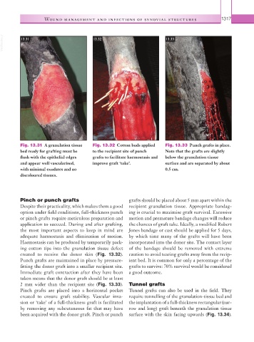
VetBooks.ir 13.31 13.32 13.33
Fig. 13.31 A granulation tissue Fig. 13.32 Cotton buds applied Fig. 13.33 Punch grafts in place.
bed ready for grafting must be to the recipient site of punch Note that the grafts are slightly
flush with the epithelial edges grafts to facilitate haemostasis and below the granulation tissue
and appear well vascularised, improve graft ‘take’. surface and are separated by about
with minimal exudates and no 0.5 cm.
discoloured tissues.
Pinch or punch grafts grafts should be placed about 5 mm apart within the
Despite their practicality, which makes them a good recipient granulation tissue. Appropriate bandag-
option under field conditions, full-thickness punch ing is crucial to maximise graft survival. Excessive
or pinch grafts require meticulous preparation and motion and premature bandage changes will reduce
application to succeed. During and after grafting, the chances of graft take. Ideally, a modified Robert
the most important aspects to keep in mind are Jones bandage or cast should be applied for 5 days,
adequate haemostasis and elimination of motion. by which time many of the grafts will have been
Haemostasis can be produced by temporarily pack- incorporated into the donor site. The contact layer
ing cotton tips into the granulation tissue defect of the bandage should be removed with extreme
created to receive the donor skin (Fig. 13.32). caution to avoid tearing grafts away from the recip-
Punch grafts are maintained in place by pressure- ient bed. It is common for only a percentage of the
fitting the donor graft into a smaller recipient site. grafts to survive: 70% survival would be considered
Immediate graft contraction after they have been a good outcome.
taken means that the donor graft should be at least
2 mm wider than the recipient site (Fig. 13.33). Tunnel grafts
Pinch grafts are placed into a horizontal pocket Tunnel grafts can also be used in the field. They
created to ensure graft stability. Vascular inva- require tunnelling of the granulation-tissue bed and
sion or ‘take’ of a full-thickness graft is facilitated the implantation of a full-thickness rectangular (nar-
by removing any subcutaneous fat that may have row and long) graft beneath the granulation tissue
been acquired with the donor graft. Pinch or punch surface with the skin facing upwards (Fig. 13.34).