
Page 1345 - Equine Clinical Medicine, Surgery and Reproduction, 2nd Edition
P. 1345
1320 CHAPTER 13
VetBooks.ir burns require a closed technique, which includes 13.37
Deep second-degree burns and third-degree
the use of an occlusive bandage, as long as there is
no infection or scab, or much exudate. The eschar
technique allows the wound to be protected by the
presence of the eschar and works best in small burnt
areas. It is not indicated for large burns or areas
where the burn may become traumatised. Since the
wound is left open, the clinician must be aware that
trauma and/or infection may occur. The third tech-
nique is called semi-open and involves the continu-
ous application of moist bandages and antibacterial
agents to the eschar. A moist dressing prevents heat
and moisture loss, protects the eschar and helps
prevent bacterial contamination and infection. The
frequent bandage changes with this technique allow
frequent wound debridement and, even though it
is time-consuming, it controls the amount of tis-
sue removed so healthy tissue is not accidentally or
excessively removed.
WOUNDS INVOLVING
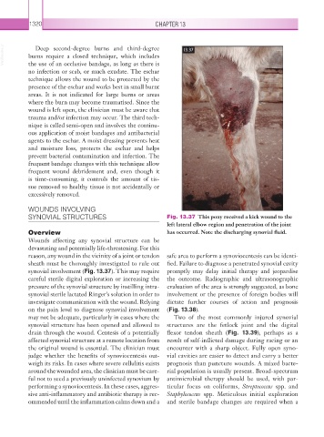
SYNOVIAL STRUCTURES Fig. 13.37 This pony received a kick wound to the
left lateral elbow region and penetration of the joint
Overview has occurred. Note the discharging synovial fluid.
Wounds affecting any synovial structure can be
devastating and potentially life-threatening. For this
reason, any wound in the vicinity of a joint or tendon safe area to perform a synoviocentesis can be identi-
sheath must be thoroughly investigated to rule out fied. Failure to diagnose a penetrated synovial cavity
synovial involvement (Fig. 13.37). This may require promptly may delay initial therapy and eopardise
j
careful sterile digital exploration or increasing the the outcome. Radiographic and ultrasonographic
pressure of the synovial structure by instilling intra- evaluation of the area is strongly suggested, as bone
synovial sterile lactated Ringer’s solution in order to involvement or the presence of foreign bodies will
investigate communication with the wound. Relying dictate further courses of action and prognosis
on the pain level to diagnose synovial involvement (Fig. 13.38).
may not be adequate, particularly in cases where the Two of the most commonly injured synovial
synovial structure has been opened and allowed to structures are the fetlock joint and the digital
drain through the wound. Centesis of a potentially flexor tendon sheath (Fig. 13.39), perhaps as a
affected synovial structure at a remote location from result of self-inflicted damage during racing or an
the original wound is essential. The clinician must encounter with a sharp object. Fully open syno-
judge whether the benefits of synoviocentesis out- vial cavities are easier to detect and carry a better
weigh its risks. In cases where severe cellulitis exists prognosis than puncture wounds. A mixed bacte-
around the wounded area, the clinician must be care- rial population is usually present. Broad-spectrum
ful not to seed a previously uninfected synovium by antimicrobial therapy should be used, with par-
performing a synoviocentesis. In these cases, aggres- ticular focus on coliforms, Streptococcus spp. and
sive anti-inflammatory and antibiotic therapy is rec- Staphylococcus spp. Meticulous initial exploration
ommended until the inflammation calms down and a and sterile bandage changes are required when a