
Page 1340 - Equine Clinical Medicine, Surgery and Reproduction, 2nd Edition
P. 1340
Wound management and infections of synovial structures 1315
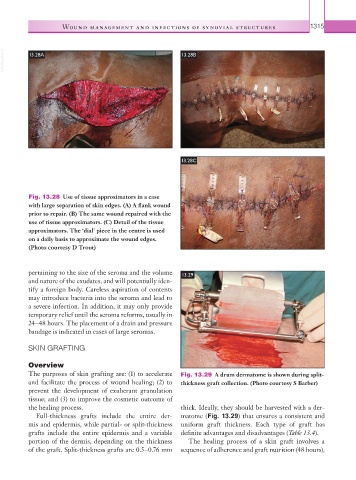
VetBooks.ir 13.28A 13.28B
13.28C
Fig. 13.28 Use of tissue approximators in a case
with large separation of skin edges. (A) A flank wound
prior to repair. (B) The same wound repaired with the
use of tissue approximators. (C) Detail of the tissue
approximators. The ‘dial’ piece in the centre is used
on a daily basis to approximate the wound edges.
(Photo courtesy D Trout)
pertaining to the size of the seroma and the volume 13.29
and nature of the exudates, and will potentially iden-
tify a foreign body. Careless aspiration of contents
may introduce bacteria into the seroma and lead to
a severe infection. In addition, it may only provide
temporary relief until the seroma reforms, usually in
24–48 hours. The placement of a drain and pressure
bandage is indicated in cases of large seromas.
SKIN GRAFTING
Overview
The purposes of skin grafting are: (1) to accelerate Fig. 13.29 A drum dermatome is shown during split-
and facilitate the process of wound healing; (2) to thickness graft collection. (Photo courtesy S Barber)
prevent the development of exuberant granulation
tissue; and (3) to improve the cosmetic outcome of
the healing process. thick. Ideally, they should be harvested with a der-
Full-thickness grafts include the entire der- matome (Fig. 13.29) that ensures a consistent and
mis and epidermis, while partial- or split-thickness uniform graft thickness. Each type of graft has
grafts include the entire epidermis and a variable definite advantages and disadvantages (Table 13.4).
portion of the dermis, depending on the thickness The healing process of a skin graft involves a
of the graft. Split-thickness grafts are 0.5–0.76 mm sequence of adherence and graft nutrition (48 hours),