
Page 1339 - Equine Clinical Medicine, Surgery and Reproduction, 2nd Edition
P. 1339
1314 CHAPTER 13
VetBooks.ir 13.26 13.27
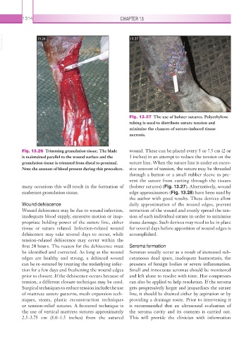
Fig. 13.27 The use of bolster sutures. Polyethylene
tubing is used to distribute suture tension and
minimise the chances of suture-induced tissue
necrosis.
Fig. 13.26 Trimming granulation tissue. The blade wound. These can be placed every 5 or 7.5 cm (2 or
is maintained parallel to the wound surface and the 3 inches) in an attempt to reduce the tension on the
granulation tissue is trimmed from distal to proximal. suture line. When the suture line is under an exces-
Note the amount of blood present during this procedure. sive amount of tension, the suture may be threaded
through a button or a small rubber sleeve to pre-
vent the suture from cutting through the tissues
many occasions this will result in the formation of (bolster sutures) (Fig. 13.27). Alternatively, wound
exuberant granulation tissue. edge approximators (Fig. 13.28) have been used by
the author with good results. These devices allow
Wound dehiscence daily approximation of the wound edges, prevent
Wound dehiscence may be due to wound infection, retraction of the wound and evenly spread the ten-
inadequate blood supply, excessive motion or inap- sion of each individual suture in order to minimise
propriate holding power of the suture line, either tissue damage. Such devices may need to be in place
tissue or suture related. Infection-related wound for several days before apposition of wound edges is
dehiscence may take several days to occur, while accomplished.
tension-related dehiscence may occur within the
first 24 hours. The reason for the dehiscence must Seroma formation
be identified and corrected. As long as the wound Seromas usually occur as a result of increased sub-
edges are healthy and strong, a dehisced wound cutaneous dead space, inadequate haemostasis, the
can be re-sutured by treating the underlying infec- presence of foreign bodies or severe inflammation.
tion for a few days and freshening the wound edges Small and innocuous seromas should be monitored
prior to closure. If the dehiscence occurs because of and left alone to resolve with time. Hot compresses
tension, a different closure technique may be used. can also be applied to help resolution. If the seroma
Surgical techniques to reduce tension include the use gets progressively larger and jeopardises the suture
of mattress suture patterns, mesh-expansion tech- line, it should be drained either by aspiration or by
niques, stents, plastic reconstruction techniques providing a drainage route. Prior to intervening it
or tension-relief sutures. A favoured technique is is recommended that an ultrasound evaluation of
the use of vertical mattress sutures approximately the seroma cavity and its contents is carried out.
2.5–3.75 cm (1.0–1.5 inches) from the sutured This will provide the clinician with information