
Page 344 - Equine Clinical Medicine, Surgery and Reproduction, 2nd Edition
P. 344
Musculoskeletal system: 1.8 Soft-tissue injuries 319
VetBooks.ir 1.616 1.617
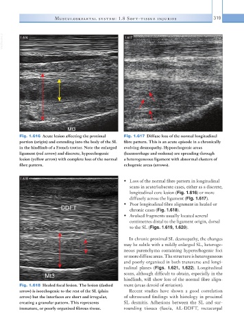
Fig. 1.616 Acute lesion affecting the proximal Fig. 1.617 Diffuse loss of the normal longitudinal
portion (origin) and extending into the body of the SL fibre pattern. This is an acute episode in a chronically
in the hindlimb of a French trotter. Note the enlarged evolving desmopathy. Hypoechogenic areas
ligament (red arrow) and discrete, hypoechogenic (haemorrhage and oedema) are spreading through
lesion (yellow arrow) with complete loss of the normal a heterogeneous ligament with abnormal clusters of
fibre pattern. echogenic areas (arrows).
1.618
• Loss of the normal fibre pattern in longitudinal
scans in acute/subacute cases, either as a discrete,
longitudinal core lesion (Fig. 1.616) or more
diffusely across the ligament (Fig. 1.617).
• Poor longitudinal fibre alignment in healed or
chronic cases (Fig. 1.618).
• Avulsed fragments usually located several
centimetres distal to the ligament origin, dorsal
to the SL (Figs. 1.619, 1.620).
In chronic proximal SL desmopathy, the changes
may be subtle with a mildly enlarged SL, heteroge-
neous parenchyma containing hyperechogenic foci
or more diffuse areas. The structure is heterogeneous
and poorly organised in both transverse and longi-
tudinal planes (Figs. 1.621, 1.622). Longitudinal
scans, although difficult to obtain, especially in the
hindlimb, will show loss of the normal fibre align-
Fig. 1.618 Healed focal lesion. The lesion (dashed ment (areas devoid of striation).
arrow) is isoechogenic to the rest of the SL (plain Recent studies have shown a good correlation
arrow) but the interfaces are short and irregular, of ultrasound findings with histology in proximal
creating a granular pattern. This represents SL desmitis. Adhesions between the SL and sur-
immature, or poorly organised fibrous tissue. rounding tissues (fascia, AL-DDFT, metacarpal